MammoDiagnost VU is Philips Healthcare's mammography workstation.
One of the biggest challenges that has plagued mammography radiologists is integrating the stand-alone mammography workstation with the multimodality workstation.
Progress in developing a truly unified multimodality reading environment has been stalled by an inability to reconcile the specific software and hardware requirements that differentiate mammography from CT, MR and general X-ray reads. The next step forward, however, is not too far off as new workstations deliver solutions that encompass the management of all types of breast and radiologic images in a single workstation. But is the implementation easier said than done?
Unique Needs of Mammography
“In breast radiology we have the issue of how are we going to read mammograms and then integrate them with other types of breast imaging that we have, such as breast ultrasound and breast MRI,” said Gillian Newstead, M.D., director of Clinical Breast Imaging at the University of Chicago.
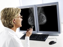
“We also have a unique situation where we require special workstations for viewing mammography images; they have to have 5 megapixel (MP) viewers. The result of this situation is that we have had lots of monitors, each doing something very specific, and we have to move from one set of monitors to another set of monitors to look at mammography, and then ultrasound and MRIs, and we can’t view everything in one integrated environment. That has been a problem that most breast radiologists face and have faced for many years. Unlike other areas of radiology where most radiologists are able to read their studies on a PACS, we haven’t been able to do that in the breast domain.”
A key differentiator between reading full-field digital mammography (FFDM) and CT or MR are the hanging and display protocols. Mammography has specific hanging protocols based upon view, laterality, patient orientation, and specialty views. The display needs to maintain a black air gap during window/center operations and inverted pixel data.
Computer-aided detection (CAD) software is also an important tool in mammography reading. The CAD application, which helps detect abnormal areas of density, mass or clusters of micro-calcifications that warrant a second look from a clinician, should come integrated in the PACS.
Another challenge involves importing images. “It has not been an easy matter to import different types of mammography images onto different vendor workstations,” said Dr. Newstead. “For example, if you have a single-modality workstation and wanted to view an image from a third-party vendor, it might not view entirely satisfactorily.”
By integrating PACS with breast imaging, this enables image fusion. With this procedure, sequentially acquired positron emission tomography (PET), for example, are fused with MR images in which the PET scan depicts glucose metabolism in cells, while the MR image localizes the tumor.
To perform web-based mammography, every workstation should use a 5MP monitor and have access to the uncompressed DICOM images. “It’s possible today to install 5MP displays everywhere, however, this is probably prohibitive from a cost perspective,” Henri “Rik” Primo, strategic relations manager, image and knowledge management, Siemens Healthcare, pointed out. “Further, many web-image distribution servers use compression algorithms. You can use lower display resolution screens to display mammography images and reports, but for referral use only, not for primary diagnosis.”
Another feature unique to mammography are BI-RADS (the Breast Imaging Reporting and Data System). BI-RADS are a collaborative effort between mammography specialists and the American College of Radiology (ACR). After reading a mammogram, radiologists assign BI-RADS scores, with 5 being highly suggestive of a malignancy and 0 representing an incomplete exam. For radiologists, it’s very useful to be able to mark up images, log them directly into BI-RADS, and store the reports directly in the RIS.
Meet the Demand
Despite the obstacles, vendors have made recent inroads into digital mammography to support the requirements of mammography in a workstation combined with other DICOM images.
At the center for Clinical Breast Imaging at the University of Chicago, radiologists struggled with importing images from a third-party vendor and having to readjust the images in order to view them properly. “The images may come over so that you can view them, but they may not line up properly or they are different sizes. There is a lot of manipulation that the radiologist has to do so that you can read them,” said Dr. Newstead.
After the department implemented MammoDiagnost VU Workstation by Philips Healthcare, doctors were able to import images easily and the automatic breast tissue alignment for images could be derived from any vendor.
“The advantage of this workstation was that once it was hooked up with our PACS, we were able to import images from different types of mammography machines. In general, when you buy a digital mammography system the vendor wants you to use their own vendor-specific mammography workstation,” said Dr. Newstead. “The MammoDiagnost VU was designed to and in fact does import quite well disparate types of mammography images and displays them in a similar fashion, which is an advantage. We are a referral center, and we get images from all over the country, so we want to import images from different types of mammography systems. One of the goals to this workstation is to do that efficiently and well.”
Because it handles a wide variety of images, the new system has helped streamline workflow. “When you are in a big center and you have images coming in from everywhere, this is a big advantage. You can view all of the images at the same workstation,” she added. “I think the display of the images is uniform. The proper auto-sizing and auto-alignment has made the reading environment much better. I like the fact that it is faster. The auto-alignment is done automatically on this workstation unlike on other workstations where it’s manual.”
Going Digital and Beyond
Similarly, the syngo MammoReport workplace by Siemens Healthcare facilitates image imports by enabling the user to retrieve old films once they have been converted or “scanned” into a digital DICOM format. At Dickinson County Healthcare System (Iron Mountain, Mich.), where doctors read 1,300 mammography exams a year, workflow efficiency is of the utmost importance. To stay competitive, the radiology department made the transition from analog to digital in October 2008 with the help of Siemens Healthcare. The solution Siemens Healthcare provided was the integration of its syngo web-enabled software with syngo MammoReport.
According to Primo, this approach adds powerful networking capabilities, including seamless RIS connectivity, multimodality viewing, third-party CAD, IHE integration, and an intuitive user interface. The PACS provides the Mammo workplace with relevant images of different modalities when comparing FFDM images with images acquired from MRI, CT, nuclear or ultrasound modalities. Each of these modalities provides different types of anatomical information in addition to FFDM.
One of the key improvements for Dickinson County Healthcare System was using RIS as a worklist. “If you have a RIS that drives the workflow and it stores the information regarding the BI-RADS and the productivity reports, this is very helpful for your annual ACR accreditation,” noted Sherry Koepp, ARRT, ARDMS, CIIP, RIS/PACS administrator, Dickinson County Healthcare System. “The digital mammo is very efficient because you can quickly pull up the patient and do the exam. It cuts down on the procedure time because before they were changing films. Plus, the feedback from our radiologists is that the image quality is excellent. The whole process is a lot more seamless and very efficient.”
Additional enhancements include mammography image viewing tools, including zoom, pan, flipping, and roaming to help better detect pathology. “Because radiologists normally don’t like too many mouse clicks, or plowing through long sequences of images to come to a diagnosis, the system is designed to provide an ergonomic keyboard, with specific shortcuts that provides an automated but user-specific workflow, guiding the user through the different workflow steps. All this shortens the time it takes for image interpretation, and ensures consistency in the process,” indicated Primo.
Koepp added, “MIP is the thing the radiologists like the most. Being able to zoom in or out on the image allows them to step through these processes at the touch of button.”
Smooth Transition
When Joint Township Hospital converted to digital, a smooth transition and physician buy-in was a priority. To achieve this they first installed the RIS/PACS by Carestream Health, allowing the radiologists to adapt to the new technology.
“About six months after the PACS arrived, we implemented our full-field digital mammography (FFDM),” explained Rob Homan, RIS/PACS/medical imaging/clinical administrator. “Already having that workstation in place, our radiologists have gotten comfortable with the flow and general toolset of PACS, so moving forward to the FFDM environment, their transition was really easy. It was just a matter of getting used to the new tools available with the mammography module. We installed it as part of our new women’s imaging department, which is a dedicated area within our medical imaging department.”
To ease into the transition, they mimicked the display protocol of a film multiloader. “Anything we could do to make it seem like they were reading in the old environment made it that much easier to transition to digital. It was nice having a PACS workstation already installed,” said Homan.
Now the radiologists at the hospital can read both mammography and switch to a stat CT from the emergency department at the same workstation.
“When a mammography case is presented, the system presents the mammography toolset. If you’re reading a chest X-ray from the emergency room, you have your basic PACS toolset. If you want to get back to reading the rest of the mammo cases for the day, you double-click on a mammography case, then get all of my mammography tools. Your CAD on and off display, your quadrant zoom and your chest wall margins [are] set,” said Homan.
Joint Township Hospital is one of the few sites in Ohio that provides molecular breast imaging (MBI), also called breast-specific gamma imaging. Because MBI will not use the same mammography toolset because it is a nuclear-medicine based exam, the administrators built the display protocol to emulate mammography.
“We are able to display those images with the mammo-all-on-one workstation,” said Homan. “We can display the MBI exam just like a mammography display so that each view is in a specific quadrant. The workstation can automatically display the comparison mammogram to compare against the MBI exam. That is a unique feature — when you have a multimodality workstation you are able to bring in a MBI image and toggle it just like a mammography case.”
More Work to Be Done
Despite these recent advances in mammography workstation technology, there is still more work to be done, Dr. Newstead reminds us.
“It is great that we are paying attention to the viewing environment because we have all of these wonderful images, but if we can’t view them efficiently, it’s a problem for the radiologist,” said Dr. Newstead. “In the future, I would like it to be fully integrated into the PACS so that we can just use routine workflow. Right now we have to push images there.”
She added, “I am really happy that industry is moving in this direction, it is something that we have been hoping for, for a long time, and I think this is an important step to really make it possible for us to view any type of mammogram efficiently and to have it be as seamless as it is.”

 June 02, 2026


 Clearance")