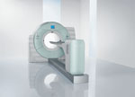
The CT system on Siemens Biograph mCT can be used as a dedicated CT and as a hybrid PET/CT.
In these challenging economic times, hospitals are seeking to maximize their investment in technology. To justify purchasing any imaging modality, such as computed tomography (CT), positron emission tomography (PET) or single photon emission computed tomography (SPECT), a facility has to have sufficient patient volume to pay for the investment.
One way to assure patient throughput covers the cost of technology to optimize the use of a modality by applying it to several clinical indications. This strategy is driving the trend toward multipurpose hybrid imaging systems with full capacity in both modalities.
Multipurpose Modalities
Nuclear medicine has made a compelling case in favor of multipurpose modalities, in particular with PET/CT and SPECT/CT technologies. The argument is that hybrid imaging benefits patient management and actually lowers health care costs.
One strategy to getting the most out of a hybrid system is to expand its clinical applications.
Philips Healthcare took the approach to expand clinical applications when it consolidated its Gemini TF time-of-flight PET imaging technologies with its Brilliance Big Bore CT. The new model, Gemini TF Big Bore PET/CT Tomograph, was designed with a large 85-cm bore. This allows patients to be positioned in the same manner for both simulation and therapy. These additional applications expanded the PET/CT’s capabilities beyond diagnosis, staging and follow-up to include therapy planning.
By consolidating procedures, hybrid modalities can serve multiple clinical purposes.
Two for One
Until recently, the CT part of the hybrid system was used just for mapping out the location of the tumor or disease. Subsequently, the CT system could only be used as an adjunct to the PET or SPECT, not as a standalone system.
The fields of radiology and oncology have emerged as optimal testing grounds for a hybrid system with full-capacity modalities.
Last year, Siemens Healthcare introduced the Biograph mCT, a molecular CT that enables health care facilities to serve both the nuclear medicine and radiology departments with one system. It achieves this duality through the integration of powerful PET and CT technologies, offering a compact system with high-definition PET, time-of-flight technology and CT configurations from 64 slices to 128 slices.
This game-changing technology allows clinicians to use a hybrid system as either a dedicated CT or as a PET/CT.
Last year, Memorial Medical Center (MMC) in Springfield, Ill., installed a Siemens Biograph mCT. The hospital replaced a PET/CT it used as a diagnostic tool for oncology management and sought to add a dedicated CT in an outpatient setting. The initial plan was to buy two separate pieces of equipment. However, when Siemens presented the Biograph mCT, the hospital decided it was more economical to buy the hybrid system.
A new PET/CT system runs in the $2 million range and a high-end CT system costs at least $1 million. “It cost $600,000-$700,000 less to buy Biograph mCT compared to buying two separate high-end systems, and we could put them both in the same room,” said Charles E. Neal, M.D., radiologist, president of Clinical Radiologists, and a physician at Memorial Medical Center, Springfield, Ill.
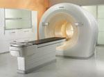
Similarly, GE Healthcare launched last year the Discovery NM/CT 670, which combines SPECT technology with multislice CT. The system is pending 510(k) clearance in the United States, but it is designed to enable clinicians to perform hybrid and standalone diagnostic CT scans on a single system.
“With the combination of advanced CT and SPECT technology available on the Discovery NM/CT 670, we will be able to optimize both our SPECT and standalone CT protocols,” said professor Ora Israel, director of nuclear medicine and PET/CT at Rambam Medical Center in Israel.
Hybrid Optimization
One of the key advantages of the hybrid model is its value proposition.
“The overall outlay of the cost is less to buy the highest end of both the PET and the CT. You can buy used PET/CT’s for less money, but the hospital wanted to offer the best available so that people wouldn’t have to drive to another location. The hospital also wanted to produce higher resolution images with the high-definition PET,” said Neal.
Another benefit of a hybrid system used for multiple applications is it rarely stands idle.
“The main thing is we didn’t buy a PET that sits empty for half a day because we use the CT for part of it,” said Neal. “Previously, nuclear medicine doctors were the only ones using the PET scanner, and then CT technologists used it only for CT scans. Now, we have a team of technologists who do the hybrid, PET/CT imaging, and the rest of the day the CT technologists do the CT imaging.”
Image Quality
Without Compromises
No clinician wants to compromise image quality for efficiency, but it is important to shorten exam times. By using two high-end imagers for a single scan, not only are scan times shorter, but image quality is superior.
The mCT at MMC is a 128-slice system that, according to Neal, performs faster than the previous CT [systems] at the hospital. Thanks to its faster scanning speed, patients do not have to hold their breath as long as they did. Less patient movement results in fewer artifacts, decreased repeat scans, and subsequently, reduces the amount of intravenous contrast agent used.
“Because we bought the high-end of both machines, we can image patients quicker on the scanner,” said Neal. “There are two issues. On the PET/CT it took 25 minutes for the patient on the table. Once the computer processed the data, it took an hour for one patient. Now it takes 10 minutes to scan each patient, so we can do two patients in an hour. It has doubled the potential throughput.”
He added, “However, we don’t have twice as many patients to scan. So what we have done is to scan our regular CT patients on the hybrid CT. We can have more patients done on the top CT because we have finished with our PET patients. We just turn it over and use it as a CT scanner the rest of the day.”
The Discovery NM/CT 670 also is designed to shorten acquisition times and improve dose management, compared to using separate, conventional SPECT and CT exams.
“We understand that a clinician does not want to compromise,” said Nathan Hermony, general manager of GE Healthcare’s nuclear medicine business. “With the Discovery NM/CT 670, we are providing clinicians the ability to discover and explore new capabilities, while continuing to perform advanced hybrid and standalone CT procedures.”
This full-featured SPECT/CT, system is engineered to reduce scan time. Bone imaging protocols, including planar whole body, 3-D SPECT and CT attenuation correction/localization, are among the most frequently performed nuclear medicine procedures. With conventional nuclear hybrid imaging, a traditional bone imaging protocol can take up to 55 minutes. With the Discovery NM/CT 670, the imaging time is reduced to as little as 16 minutes. Shorter scan times result in less patient movement, which minimizes artifacts in the images.
Improved Diagnosis
The faster acquisition times on both the mCT and NM/CT 670 are designed to improve image quality and clinical diagnosis.
“We can see things easier now with the new scanner compared to the old one. Previously with the PET/CT, they put a low-end CT on it to just show the location in the body, but the CT didn’t give you the full detail of a regular CT,” said Neal. “Now with both high-end PET and CT, we can see quality images in the same scan. It helps in diagnosis to tell more precisely whether it’s positive or negative on the PET scan.”
He added, “If there was a previous PET scan, we can see whether it has gotten better, worse or is the same. As a result, we have a better understanding of the overall response of the tumor to the treatment.”
With hybrid imaging, a scan on one of the modalities may be more accurate than one acquired simultaneously on the other system. For example, the CT image may show a tumor is shrinking, while the PET exam indicates it is expanding.
In a recent case, Neal treated a patient who had lymphoma. On the CT, all of the tumor sites showed the tumor had reduced in size, which told the oncologist the patient was responding well. But two sites on the PET scan appeared worse.
“If the doctor hadn’t changed the course of treatment based on the PET data, the patient was not going to have a good outcome,” Neal said. “In that case, what the hybrid imaging showed you was something had to be changed in the treatment regimen.”
Ora concurs that the hybrid does a better job of characterizing the state of the tumor.
“The technology has the ability to improve localization and overall clinical confidence in routine scans and research applications,” said Ora.
As economic pressures drive down capital expenditures in heath care, hospitals will increasingly seek hybrid solutions to get more bang for the buck.
For More Information
www.gehealthcare.com
www.medical.philips.com
www.siemens.com/healthcare

 July 09, 2026


