Early detection is an important and valuable tool in the continuing fight against breast cancer. Research has found that cancers discovered during early screening exams are often smaller in size and still confined to the breast compared to those found later. When considering these two factors, the size and spread of breast cancer — the most important predictors of prognosis for women with the disease — early detection has become a focal point in cancer care. As a result, the American Cancer Society (ACS) has established guidelines that recommend women over 40 undergo annual screening mammograms.
While clinical evidence supporting the need for annual mammograms, especially in the younger population, is still developing, mammography itself continues to prove to be a valuable tool in the fight against breast cancer.
The current gold standard in breast cancer screening is 2-D mammography. Although 2-D full-field digital mammography (FFDM) is successful in identifying a large number of cancers, this method has its limitations. A traditional 2-D mammography study consists of two images per breast — caniocaudal (CC) and mediolateral-oblique (MLO). These flat images can be challenging to interpret as tissues may overlap and obscure cancerous areas.
However, digital breast tomosynthesis (DBT) has found a way to help overcome this challenge. During DBT, the mammography system rotates in an arc around the breast while taking a series of images. On average, a total of 10–20 low-dose images are acquired. These images are then reconstructed to create a computed tomography (CT)-like study that enables the radiologist to view the breast in smaller slices, typically around 1 mm thick. By utilizing DBT, the radiologist can see the breast in segments and create a 3-D rendering. This additional imagery increases physicians’ ability to view cancers that would otherwise appear obscured in traditional 2-D mammography, and decreases the number of biopsies and recall rates.
Current State of the Market
Because DBT is a new technology, the U.S. Food and Drug Administration (FDA) requires manufacturers to go through a premarket approval (PMA) process, the most stringent type of device marketing application, rather than the traditional 510(k) clearance. Since there is no substantially equivalent predicate device, the PMA application process can be tedious and time-consuming, as it requires vendors to prove both the safety and effectiveness of their device. PMA applications almost always require the submission of documented clinical research data, which takes time to compile.
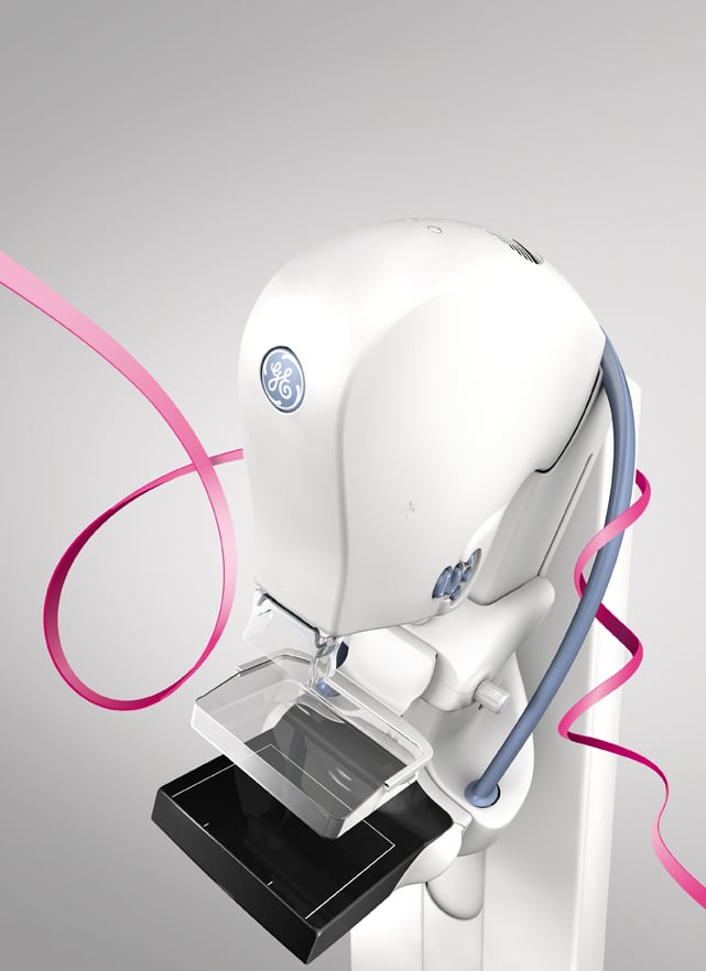
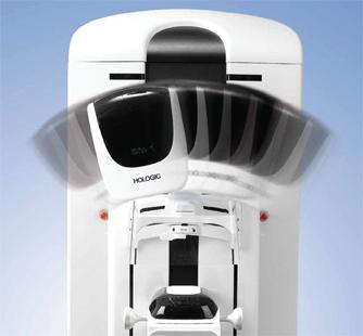
Hologic and GE Healthcare are currently the only two vendors that have received FDA approval for their technology in the United States. Hologic’s Selenia Dimensions tomosynthesis system has been available in the United States since 2011, while GE Healthcare’s SenoClaire 3D tomosynthesis system was approved in September. Siemens Healthcare only recently submitted its PMA applications to the FDA in June 2014.
Currently, all three vendors offer DBT solutions outside of the United States, which helps to create a competitive market that provides facilities with system choices and helps drive down the cost of investing in a DBT-capable system. How exactly GE’s recent approval in the United States will change the market remains to be seen, although the presence of a second vendor will offer some much needed competition and provide hospitals with better negotiation leverage.
System Pricing
Hologic’s Selenia Dimensions 2-D system, which costs around $300,000 on average, is upgradeable to 3-D for $125,000–150,000. At the time of this publication, pricing for GE Healthcare’s SenoClaire 3D tomosynthesis system has not been released. (See Table 1.)
Reimbursement Expectations
Currently, there is no dedicated reimbursement code for 3-D tomosynthesis. Early adopters of this technology have utilized the existing reimbursement codes for the 2-D portion of the exam, but are unable to bill for the additional 3-D imaging component. Facilities looking to recoup this lost revenue have found some success in filing for additional reimbursement under CPT 76499 (miscellaneous radiographic procedure) although reimbursement through this route is not always guaranteed. Some facilities will charge the patient a flat fee, usually around $50–75, while others who feel the benefits of the technology are worth the additional expense will absorb the cost themselves.
However, concerns over the lack of reimbursement may change as the American Medical Association (AMA) CPT Editorial Panel has approved three new Category I CPT codes for breast tomosynthesis that will be effective beginning Jan. 1, 2015. Although approval of these codes does not guarantee the Centers for Medicare & Medicaid Services (CMS) will follow up with an assigned professional payment rate and technical code for breast tomosynthesis, it is an encouraging first step toward independent reimbursement from both CMS and private insurance. It is difficult to determine if and when CMS intends to address the issue, but once the Medicare Physician Fee Schedule Final Rule is issued by the CMS sometime in November, the outlook of reimbursement for DBT will become clearer.
Regardless of what the future may hold, one thing remains true — reimbursement continues to play a role in a facility’s decision to invest in a tomosynthesis-capable digital mammography system. Some facilities, especially smaller ones, are waiting for reimbursement before their administrators will allow them to purchase DBT. Others that currently utilize DBT are concerned the dedicated DBT reimbursement may be less than what they are currently receiving, typically through charging the additional $50–75 fee.
Other Considerations
Price and reimbursement are not the only factors to take into consideration when looking to invest in DBT technology. Providers must take into account changes in radiation dose, the larger file size, storage needs and required software/hardware upgrades when transitioning to 3-D tomosynthesis. These considerations can be costly, but the measurable benefits of offering DBT include the ability to maintain the competitive edge when competing for patients and referring physicians, positive customer feedback and compelling clinical evidence.
Radiation Dose. When Hologic first received FDA approval for its Selenia Dimensions tomo system, patient dose was a huge topic of conversation. Acquiring a tomosynthesis study of approximately 10–20 low-dose images in addition to the 2-D exam exposes the patient to a slightly higher radiation dose. Since then, Hologic has released its C-View option that allows the tomosynthesis exam to be reconstructed into 2-D images, eliminating the need for additional exposures. It remains to be seen how other vendors will handle this issue, but it is something to be aware of.
Larger File Size. Storage space is another topic for consideration. A 2-D FFDM study contains a total of four images. Varying with breast size, DBT studies can contain anywhere from 50–100 reconstructed images per breast. Consideration should be taken to ensure the picture archiving and communication system (PACS) is equipped to handle these massive image files which must be stored in their original format. Because of the increased number of images, the amount of time required for a radiologist to read tomosynthesis studies is longer than the traditional 2-D FFDM. Additional training will be necessary to ensure that all staff is knowledgeable on the differences in DBT imaging.
System Upgrades. Workstations must be reviewed to ensure they contain the proper software to view tomosynthesis studies. Hardware upgrades are sometimes necessary to support the larger file size associated with DBT images.
Maintaining the Competitive Edge. While a recent Gallup poll showed that most Americans are satisfied with their quality of care (82 percent),1 the information age is changing how patients seek out this care . They are often better informed about their options and have a better understanding of the quality of care a provider can offer than ever before. Media stories and the Internet have made it exceptionally easy for patients to learn about advantages of DBT systems. As this trend continues and patients become increasingly adept at choosing their providers, hospitals will face the added challenge of competing for patients. This is a critical issue since volume is a key factor for the success of any service line.
Furthermore, when referring physicians are aware DBT is available at a certain facility it breeds confidence in sending patients to that location. Gaining more referrals, especially in an urban area where multiple hospitals compete for the same patients, can often be a huge challenge.
Customer Feedback. While there are a lot of factors to consider when purchasing this technology, customers have reported that tomosynthesis is the best investment they have ever made. User feedback from MD Buyline’s database reports that DBT has increased business, increased cancer detection and decreased callbacks by 25-32 percent. In addition, Hologic’s Selenia Dimensions system has a faster acquisition time in comparison to the previous generation Selenia system, minimizing patient discomfort. Customers also report the Selenia Dimensions system has improved image quality and provides a sharper picture than the Selenia system.
Clinical Evidence. Numerous studies have been conducted to evaluate the benefits of DBT technology. One recent study by the Journal of the American Medical Association (JAMA) evaluated 454,850 examinations across 13 facilities to determine if there was truly an advantage to utilizing tomosynthesis in breast screenings.2 This study concluded that tomosynthesis images provided a 41 percent increase in the detection of invasive breast cancers and a 15 percent drop in recalls for additional imaging.
Another study published in Radiology compared the recall and cancer detection rates of tomosynthesis plus conventional digital mammography to those of conventional digital mammography alone for 13,158 patients.3 This study found that tomosynthesis could particularly benefit women with dense breasts and women younger than 50.
What is the significance of these results? Not only are more cancers being detected with the use of tomosynthesis, but overall diagnostic costs are reduced because of the drop in recalls for additional imaging. With the size and spread of breast cancer being the most important predictors of prognosis for women with the disease, finding these cancers sooner, when they are smaller and still confined to breast tissue, can greatly improve patients’ prognosis.
Future Outlook of the Market
The clinical evidence surrounding tomosynthesis continues to grow, and supports its use as a tool for early breast cancer detection. As studies are able to better identify those patients who will most greatly benefit from its use, this will likely influence and drive reimbursement. A large number of facilities have already adopted this technology. Expect to see more investments in DBT systems if CMS approves the addition of dedicated reimbursement codes and more vendors enter the market.
Rachael Bennett joined MD Buyline in 2008 with seven years of clinical experience in the medical field. She is the primary clinical analyst for linear accelerators, stereotactic radiosurgery systems, mammography systems, biopsy systems and other radiation oncology and women’s health capital equipment codes. She currently holds registries as both a radiographer and radiation therapist through the American Registry of Radiologic Technologists (ARRT).
Katie Regan joined MD Buyline in 2013, bringing with her seven years of clinical research experience. At MD Buyline, Regan is responsible for all clinical, financial and general healthcare publishing projects. Her work has been published in hfm Magazine, Becker’s Hospital Review, ITN and DOTmed.
References:
1 www.gallup.com/poll/159455/americans-satisfaction-health-coverage-slips-slightly.aspx. Accessed Aug. 22, 2014.
2 www.ncbi.nlm.nih.gov/pubmed/25058084. Accessed Aug. 22, 2014.
3 www.ncbi.nlm.nih.gov/pubmed/23901124. Accessed Aug. 22, 2014.

 July 02, 2026


