SpeedSuite pairs the high-speed Fuji Cassetteless imaging readers with X-ray equipment.
A 30-year-old woman arrives in the emergency room with “abdominal cramping and vaginal bleeding. A physical examination reveals a palpable left adnexal mass. She has a positive pregnancy test with beta-HCG of 14,000 IU/L. The emergency department (ED) physician considers magnetic resonance imaging (MRI) of the pelvis, yet it is expensive and not the initial study of choice. An endovaginal ultrasound shows there is no intrauterine pregnancy, that the fetal heart rate is 139 bpm, and there is a small amount of free fluid visible in the cul-de-sac.”1 The question is, what imaging device will most expeditiously and accurately indicate that these findings are diagnostic of a left ectopic pregnancy?
This is a real-life case from the Brigham and Women’s Hospital, Harvard Medical School, where the ED physician had to quickly choose from among a complex set of imaging modalities, keeping in mind managed care’s emphasis on cost-effective allocation of resources, to accurately diagnose the patient. For years, hospital ED’s have relied on ultrasound, computed radiography (CR) and digital radiography (DR) to triage patients, however, as more hospitals large and small are equipped with CT and MRI scanners, ED physicians have access to more sophisticated devices that are increasingly used to diagnosis and triage trauma patients. Even multislice CT angiography may become the new standard in the ED for patients with systems for serious conditions such as acute myocardial infarction or coronary embolism.
Ultrasound Showing Vital Signs
While ultrasound has long been a staple in the ED, its importance in triaging patients is growing stronger nationwide. “The portable ultrasound is used for central intravenous line placement, thoracentesis, paracentesis and peripheral arterial pulse location. It is the only imaging device we currently have in the ER,” said C.E. Ballard, M.D., an ED physician at Baptist Hospital, Little Rock, AR.
At Baptist Hospital, as in a growing number of facilities, ultrasound is key to expediting patients, and across the U.S. “diagnostic ultrasound utilization has grown steadily,” according to a survey by IMV Medical Information Division taken on 2,975 hospital-based general ultrasound or radiology departments. The research indicated that “[f]or hospitals with 200 plus beds, patient exams grew 41 percent from 13.1 million in 1998 to 18.5 million in 2005, for an annual average growth rate of five percent per year.” Ironically, a recently released study by Yale University School of Medicine revealed that two thirds of emergency departments do not currently have access to ultrasound for emergency physician use.
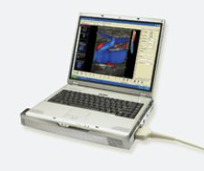
With heightened interest in ultrasound for the ED, manufacturers are designing compact units for trauma units. GE Healthcare just launched LOGIQ e, a member of GE’s Compact Series of ultrasound systems, which rapidly produces high image quality to support real-time clinical decisions in emergency settings. “We’ve designed the LOGIQ e to help extend the reach and utility of advanced ultrasound imaging to emergency patient care – providing emergency physicians with valuable diagnostic information to support life-critical situations such as trauma, abdominal aortic aneurysm or heart conditions,” said Omar Ishrak, president and CEO of GE Healthcare’s Clinical Systems division. GE has also introduced the Voluson i, a laptop-sized ultrasound system with 4-D imaging.
Another new portable ultrasound system is the Terason t3000 Ultrasound System, which combines a front-end Fusion Processor with powerful PC-based back-end data processing that runs on Windows and can be carried as a cart-based system.
Ultrasound is also showing renewed vital signs as a patient monitoring and tracking tool. As part of the Scalable Medical Alert and Response Technology (SMART) project, Brigham and Women’s Hospital, Harvard Medical School, in collaboration with the Massachusetts Institute of Technology (MIT), is testing ultrasound as a means of monitoring the location and vital signs of patients in the ED.
Instead of using the RFID indoor positioning system already installed at Brigham and Women, the hospital decided to link its ultrasound system with battery-powered tags to transmit 20-40 kHz of acoustic signals to receivers placed inside fanny packs that patients wear while in the ED waiting room. Once the patient moves, it triggers the tag, provided by Sonitor Technologies, to send a unique signal to the receivers. Algorithms read the signals before transmitting the IDs to a central server via a wireless LAN. From a lead attached to a patient’s fingertip, the tag monitors the patients’ heart and blood-oxygen saturation levels. The tag forwards that data to a PDA, also inside the fanny pack, which sends it to staff PDAs over a Wi-Fi network. This allows clinicians to not only monitor the vital signs of patients complaining of chest pains or shortness of breatbut it also facilitates patient localization and throughput in busy emergency departments, where time is of the essence.
Cassette Versus Cassette-Less
When dealing with trauma patients in the emergency room, speed and accurate diagnosis are critical. CR and DR devices rapidly provide quality digital X-ray images, while delivering less radiation than devices such as CT or PET, and cost a lot less.
“Emergency rooms today are overwhelmed with patients, so you have to handle more patients in less time. With an image every 20 seconds, you can see the workflow advantage of the DR. DR is faster and offers superior image quality. However, CR is less expensive and can service multiple X-ray rooms. CR is also helpful because you can put the CR cassette under a patient to take an image, which minimizes patient movement,” explained Phil Bullington, DR digital sales specialist, Kodak’s Health Group.
Manufacturers are combining the attributes of both systems to create models with rapid, high-quality image generation, typical of DR, that are portable and flexible for imaging at difficult angles, all at a CR price point.
As part of Konica Minolta’s campaign to produce CR imaging that is as good as DR, it has launched the REGIUS 370 upright CR system, equipped with new plate technology designed to increase detective quantum efficiency (DQE) to near-DR quality. It has also upgraded its Xpress CR, which now takes just 24 seconds to preview images and 40 seconds cycle time.
The burning question today, however, is not CR versus DR but cassette versus cassette-less, according to Penny Maier, Fuji’s national marketing manager, Digital X-ray. “We use a type of storage phosphor as a detector device because when hospitals are looking to go to DR they want to know if it uses cassettes or not. We very much see a combination of both units, which don’t require cassettes,” said Maier. Cassetteless CR systems combine image capture, reading and image upload in one device itself, making cassetteless CR systems similar to DR. Following this line of thought, Fuji has plans later this year to introduce the Carbon X CR unit with comparable image quality to a DR and with a maximum per hour throughput of 72 images.
Implementing a hybrid approach, Kodak will soon launch Kodak Point of Care CR ITX 560, a mobile digital CR system for the ER or ICU. “The X-ray system and CR system are integrated and both mounted on the cart. It helps with workflow and productivity because you can see the image right there and determine if there is a need for a retake,” said Eileen Heizyk, worldwide marketing manager in Digital Capture, Kodak’s Health Group. “One of the major drivers is cost and this is less than a DR system.” While CR and DR are appropriate for many trauma cases, when it comes to diagnosing critical diseases presented to the ED they are limited.
Triple Rule Out in the ED
Even at smaller sites, such as the 248-bed Baptist Hospital, it is becoming common to use CT for trauma patients. The ED at Baptist, indicated Ballard, “uses CT for trauma, cerebrovascular accidents, pulmonary embolus, intrabdominal, intrathoracic injuries/diseases and obstetrical, gynecological and urological evaluation.”
CT is also gaining importance in the ED for diagnosing heart disease. Chest pain is a common complaint among patients, yet failure to diagnose acute myocardial infarction in the trauma unit may occur in 20 percent of the cases, according to some estimates. Among other serious conditions occasionally overlooked in the ED are pulmonary embolism, stroke, meningitis and appendicitis. Standard protocol for chest pain is to perform an echocardiogram (ECG), the concern is, however, that physicians may rely too heavily on this test, which does not rule out a heart attack if results are normal.
In a recent study presented at the 2006 American College of Cardiology (ACC), physicians compared patients with acute chest pain who were immediately examined on a 64-slice CT with those who received a standard of care evaluation, including serial cardiac enzymes, ECGs and sestamibi rest-stress nuclear scanning. The results showed that CT angiography (CTA) rapidly and definitely excluded coronary artery disease as the cause of acute chest pain in less time and at a lower cost than stress imaging.
“The emergency room is an area where CTA could be used to its advantage. Many patients come in without obvious systems or with chest pain. The first thing we try to exclude is coronary disease. If you could do a CTA on that group of patients and exclude coronary disease, you could send those patients home early,” said James Carr, M.D., Northwestern Memorial Hospital (NMH), Radiology. A year ago, NMH installed a 64-slice CT scanner, Siemens’ SOMATOM Sensation 64; however, Dr. Carr explained that the hospital does not routinely use it for cardiac patients in the ED.
“One of the advantages of 64-slice CTA is its ability to visualize coronary arteries, and if the coronaries are normal, that effectively excludes coronary disease,” said Dr. Carr. Accordingly, he believes CTA will give a very high specificity for disease, meaning that if the study is normal nearly 98 times out of 100, the patient definitely does not have coronary artery disease. “That same scan, however, does not rule out aortic dissection and pulmonary embolus because the technique is different,” noted Dr. Carr. “To be able to rule out all three on the same scan can be challenging because there are slightly different techniques. It is known as the ‘triple rule out’ study. We are going to investigate the triple rule out study in the context of the ER, so we’re working closely with ER physicians and cardiology physicians. We will do CTA to rule out coronary disease.” To do that, NMH will install a new 64-slice scanner in the ED in the next few months.
Dr. Carr anticipates that CT, as well as MR, will become increasingly important in the ED for excluding coronary disease. He added, “It’s a huge financial savings if you can discharge patients immediately, and free up space in the ER.” Multi-slice CT dedicated to the ED may be just what the doctor ordered.
1. Finding-the-Path. http://brighamrad.harvard.edu/education/online/ftp/FTP.html. Department of Radiology, Brigham and Women's Hospital Harvard Medical School.


 June 18, 2026


