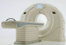
Core 320 will measure the performance of Toshibas Aquilion ONE for CCTA with myocardial perfusion against SPECT and invasive angiography.
It’s no secret many clinicians have predicted coronary CT angiography (CCTA) would replace invasive angiography for the detection of coronary artery disease for low- and medium-risk patients.
The final results of the first multicenter, multinational CORE 64 trial — a trial that compared 64-slice CTA against invasive angiography — published in the New England Journal of Medicine in November 2008, offered compelling evidence that CTA wasn’t ready to replace the gold standard invasive angiography just yet. The results of the trial showed the negative and positive predictive values for the test to be 83 percent and 91 percent, respectively, results that were somewhat disappointing to the researchers.
As announced at RSNA 2008, the new Toshiba-sponsored CORE 320 trial will compare the effectiveness of the Aquilion ONE 320-detector row dynamic volume CT to SPECT in an attempt to determine whether the combination of CTA and myocardial perfusion can identify coronary stenoses that are less than or equal to 50 percent of Quantitative Coronary Analysis (QCA) and correspond to a SPECT perfusion defect.
“CORE 320 is designed to evaluate CT in a new way,” said Frank Rybicki, M.D., director of cardiac CT and vascular CT, MRI, Brigham and Women’s Hospital, MA. “CT, in the heart, gives morphology data; anatomy and coronary imaging. It has pretty good spatial resolution and pretty good temporal resolution, and of course, it’s noninvasive. In many cases at the end of the CT scan, there are still several unanswered questions.
“The major hypothesis of CORE 320 is that the [perfusion] data can somehow be additive to morphology. CORE 320 is trying to test the value of the additional information that you can get beyond morphology.”
Both Dr. Rybicki and Rich George, M.D., assistant professor of medicine, division of cardiology, Johns Hopkins University, said CT perfusion imaging on the Aquilion ONE has shown promise in single-center studies, but only studies like CORE 320 will offer the proof that CT perfusion imaging really adds to CCTA.
“Everyone that is involved in this has high expectations,” Dr. Rybicki said. “Everyone in the community is pulling for a CT system, like a wide area detector system, to do good functional imaging and do good perfusion. Having all those expectations is wonderful, but you really have to roll up your sleeves in terms of proving it. You can do all you want in a small, single-center study, but the proof is in the pudding. You’ve got to have a study that is multicentered and multinational to really see how these technologies are going to make a global difference.”
As compared to 64-slice CT, the Aquilion ONE gives better morphological data, many clinicians say. Dr. George noted the Aquilion ONE offers faster scan times, lower radiation dose, eliminates more artifacts and decreases contrast dose from 90-100 cc to around 60 cc.
The big question for many clinicians is how the perfusion component of the Aquilion ONE will perform against SPECT, the recognized gold standard for myocardial perfusion imaging.
“The perfusion component is still in evaluation,” Dr. Rybicki said. “The perfusion images I’ve seen to date have very good technical quality, but perfusion CT is still not a clinical tool. We’re trying to make it a clinical tool. It has to be emphasized that using any scanner, including the Aquilion ONE, for perfusion and relying on the presence or absence of a perfusion abnormality is a research endeavor. That has to be stressed.
“In SPECT, it is fundamentally different. You introduce a radioisotope that goes to the heart, then radiation is emitted from the heart out and detected by a detector system. CT is totally different. You’re putting the radiation outside through and then detecting on a system. It’s not so much that it is lacking in that it’s fundamentally different and fundamentally not validated. It’s exciting, but it’s not validated.”
The performance of CT perfusion will be measured against SPECT in a similar fashion as CCTA is measured against the invasive angiography, according to Dr. Rybicki.
“You partition the heart muscle into segments, then you do your analysis that is very similar to CT,” said Dr. Rybicki. “If you see an abnormality in perfusion on CT, then you can go and look at the same abnormality using the same partition — the same myocardial segment — with SPECT.”
Dr. Rybicki said these perfusion segments help to determine the best course of treatment.
“What these bull's eye or polar maps do is segment the heart muscle designed to do this type of matching,” said Dr. Rybicki. “It’s really important to segment the heart muscle. If someone has ischemia, there are things you can do. You can manage that patient with medicine. You can do a catheter-based intervention and put in balloons and stents. Or, you can send a patient to bypass. The reason we do perfusion or SPECT is to separate patients into those categories. That is one of the great things that nuclear medicine and SPECT helps us to do. Perfusion is one piece of the puzzle used to make that distinction, to help determine the management of patients. If perfusion says the piece of heart is not being properly perfused, but it’s not dead, those patients can generally benefit from bypass. If the patient has a big perfusion deficit, and all the tissue is dead (like they had a heart attack), no amount of intervention is going to help. It would be a waste of time and money to send that patient to the OR.
“This is a tool to more accurately determine which patients go into the optimal treatment arm. To do that now, you need several things: you need images of the coronaries, you need the perfusion, you need so-called viability to determine whether the thing is dead or not. People have always thought that one modality can do all of this stuff. It hasn’t come to that yet. We’ll probably get there someday. This stuff works in increments. The 320 is a big new increment.”
Drs. George and Rybicki believe that, in all probability, CCTA with perfusion imaging will still be most beneficial for low- to medium-risk patients, as the scan still has problems imaging the heavily calcified arteries.
“[The problem of heavily calcified arteries] is not going to change with the 320-detector,” Dr. Rybicki said. “One thing that will be really nice is that if you have an artery that is heavily calcified and you have multiple lesions where there are small segments of disease, having a normal perfusion CT, if it were to validate that, would be very useful. What this would mean is that if you had all these calcifications, you still had good perfusion. That would be a real benefit. That might target more the high-risk patients. But, again, that is really a hypothesis at this point.”

 June 18, 2026


