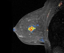
Confirma's next-generation solution includes improved 3D renderings and tools for morphology reporting.
Much controversy has surrounded the best methods of screening and detecting breast cancer as it pertains to the use of computer-aided detection (CAD).
The credibility of CAD was shaken in 2007 with an article published in the New England Journal of Medicine that found that computer-aided detection (CAD) did not improve the diagnostic accuracy of the sites using it. This report led insurance companies to threaten to stop reimbursement for CAD for breast. In a statement in September 2007 by UnitedHealthcare, the insurance mogul said, “A review of recent clinical evidence in published peer-reviewed medical literature leads us to conclude that clinical evidence does not support the use of computer-aided detection for mammography.”
However, after the initial shock and with the backing of numerous studies that support clinical benefits of CAD, insurers reversed their position and continued their coverage of CAD for breast.
One of the more recent breast studies led by Lilian Wang, M.D., at the Seattle Cancer Care Alliance and the University of Washington suggests that the CADstream system’s patented “worst curve” algorithm for analyzing kinetics may assist in determining malignancy. The study found that the most suspicious curve identified by CADstream was significantly different between benign and malignant lesions. This finding falls directly in line with the American College of Radiology’s Breast Imaging Reporting and Database System (BI-RADS) Atlas for breast MRI to report the “worst looking curve.”
Another study using Hologic’s R2 ImageChecker CAD tool found that CAD correctly marked 99 of 103 consecutive asymptomatic breast cancers, which is a 96.1 percent detection rate and a rate of 1.8 false marks per patient.
Consistency Outweighs Skepticism
Despite inconsistent findings, which may leave physicians questioning whether to use CAD as a diagnostic tool in screening for or staging women with breast cancer, many physicians continue to turn to CAD time and time again.
For David Gruen, M.D., radiologist at Norwalk Hospital in Norwalk, CT, CAD is a necessity for women who are undergoing breast cancer staging, who are pre- or perimenopausal, who have dense breast tissue or who have a family history of breast cancer. Dr. Gruen uses Confirma’s CADstream system for magnetic resonance imaging (MRI) studies, a solution developed with specific tools for each application in order to streamline workflow and standardize study analysis. Dr. Gruen asserted, “I really believe that using CADstream or any CAD package is almost a necessity for breast MR, which generates about 2,000 images per patient. CAD is really computer-aided visualization, and it more easily and rapidly identifies lesions to determine if they are worrisome or ‘leave them alone’ lesions.” He added, “The kinetics of the lesion or multiple lesions pose challenges in how to identify one that is suspicious from a forest that are insignificant. [CAD] improves accuracy and decreases false positives. We are biopsying fewer unnecessary lesions.”
In comparing his use of CADstream to other CAD products that he used in the past, Dr. Gruen explained, “The breast, chart and curve all looked different. CADstream creates internal consistency. One of my partners can get the same data tomorrow or next month. It allows for inter-observer inter-reliability.”
He mostly uses CADstream to stage women who have recently been diagnosed with breast cancer. For this type of patient, CAD is useful in that it determines whether the cancer is multifocal or multicentric and better dictates the plan of treatment, according to Dr. Gruen.
Streamlining Workflow and Throughput
Streamlined workflow from the MR system to the workstation where the CAD resides and assists the radiologist with the read is essential for optimizing the technology.
DynaCAD by Invivo is a digital imaging workstation coupled with computer-aided detection (CAD) tools for performing real-time image analysis of MR data and planning of interventional procedure planning. It is designed to provide radiologists with a comprehensive solution for improved workflow.
In addition to improving the accuracy of interpreting scans, Dr. Gruen purports that using CADstream also improves his efficiency. “It is almost cruel and unusual punishment to say you have to drive 60 miles in order to get a biopsy. If we do a breast MRI today, we can do a breast biopsy tomorrow. It improves our efficiency.” In a clinic that performs about eight to 15 breast MRI’s a day, CADstream shortens interpretation time and improves throughput, while increasing accuracy.
Other benefits of Confirma’s CADstream include its ability to integrate with existing equipment and provide access to studies everywhere on the network. Physicians in multiple locations can securely access CADstream from existing computers through CADalyst, it’s Web-based user interface. CADstream comprises fast, automated processing, comprehensive kinetic and morphology tools and advanced surgical planning tools.
Another CAD for breast product is AuroraCAD. The Aurora 1.5T Dedicated Breast MRI System with Bilateral 3-D SpiralRODEO is the only FDA cleared, fully integrated MRI system designed specifically for breast imaging. The Aurora System uses SpiralRODEO, which creates an elliptical, homogenous field of view, providing coverage of both breasts, chest wall and axillae in a single bilateral scan, reportedly without compromise in contrast or resolution. This acquisition technology provides even higher resolution, while dramatically decreasing scan time for better dynamics.
Contrast Creates a Clearer Picture
According to Pamela Donlan, M.D., radiologist at Breast Cancer Specialists in Atlanta, GA, using Aurora’s CAD improves her efficiency and accuracy in interpreting scans. She said, “What I’ve primarily noticed is that everything is integrated when the case comes over to the scanner. It allowed me to quickly establish a scan pattern. We’re getting thousands of images, and we not only have to look at images but we also have to look at acquired images, fraction images, 3D images and kinetic analysis. If I had to do this on my own, I wouldn’t get through very many cases; I couldn’t read MR without it. It automates a lot of data and presents it in a consistent, standard fashion so that once I start reading my cases, I can be extremely efficient in getting through a lot of work.” Regarding her ability to be more accurate in reading scans, she said, “One of the things that really helps is to standardize the information that radiologists are presented with and minimizes false positives.”
The Aurora 1.5T Dedicated Breast MRI System also boasts that the design provides the largest access to breasts and the feet-first entry minimizes feelings of claustrophobia. This type of comfort also reduces the motion of the patient, thereby reducing motion artifacts in the scan.
Howard Lee, M.D., a radiologist at North East Radiology, uses iCAD’s Second Look Digital, which combines hardware from GE Healthcare, Hologic, IMS Giotto and Siemens Healthcare to automatically identify and clearly mark suspicious cancers without obscuring the region of interest. At North East Radiology clinicians have been using iCAD for over five years and perform 20,000 – 25,000 mammograms a year. According to Dr. Lee, the solution allows users to detect lesions under 5 mm in size. “I have been impressed with the microcalcifications. There have been very few cases where there was a cancer proven by biopsy that did not show up,” said Dr. Lee. “The goal is to not have a high specificity rate. What you need is an acceptable false-positive rate, but not a low false-positive rate.”
CAD Solutions Coming Off the Assembly Line
Although adoption of CAD for breast may have obstacles to overcome due to the controversy regarding its effectiveness, CAD has still become a necessity for a growing number of radiologists to feel more confident in their interpretation of scans. Furthermore, the developments in CAD have not slowed down. In fact, more plans are underway to continue the growth of CAD. One exciting venture lies ahead for the Penrad Mammography Information System and CADstream which plan to integrate the two technologies in order to address workflow needs for breast imagers.
“The integration between the Penrad Mammography Information System and CADstream streamlines breast MRI reporting,” said Wayne Wager, president and CEO, Confirma.
CADstream for breast is designed to automate analysis, reporting and interventional planning of studies and promotes standardization with the incorporation of BI-RADS, which guides the breast cancer diagnostic routine and decision-making process. Confirma’s next-generation version of CADstream for breast MRI, launched at RSNA 2007, includes a new customizable BI-RADS-centric user interface that accommodates a variety of user experience levels. Additional CADstream enhancements include therapeutic monitoring capabilities, improved 3D renderings and tools for morphology reporting.
“The Penrad-CADstream integration…addresses a critical workflow need for breast imagers, which is a large segment of the breast MRI market,” Wager noted. “The ability to standardize and streamline reporting will further expand adoption of breast MRI.”
Demand for breast MRI studies has recently gained momentum with clinical studies supporting breast MRI for improved breast cancer detection and the American Cancer Society’s March 2007 recommendation that women with a 20 to 25 percent or greater lifetime risk of breast cancer should undergo an annual MRI in addition to mammography. Continued advances in CAD technology should bolster clinicians’ confidence in CAD for breast MRI and drive demand for this application.

 July 02, 2026


