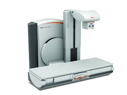
Image courtesy of Carestream
The U.S. Food and Drug Administration (FDA) defines radiographic/fluoroscopy (R/F) as a type of medical imaging that shows a continuous X-ray image on a monitor, much like an X-ray movie. During a fluoroscopy procedure, an X-ray beam is passed through the body. The image is transmitted to a monitor so the movement of a body part or of an instrument or contrast agent through the body can be seen in detail.
Fluoroscopy procedures are performed to help diagnose disease, or to guide physicians during certain treatment procedures. In years past, a dedicated fluoroscopy room was a must-have for any hospital. However, due to a shift toward other imaging modalities and changes in reimbursement, radiographic/fluoroscopy schedules are no longer full, making it inefficient and less cost-effective for hospitals to invest in a dedicated room. For this reason, many hospitals are now looking for systems that can serve multiple imaging rolls for R/F, digital radiography (DR) X-ray and in some cases for simple image-guided procedures.
Current Applications
Carestream’s DRX-Excel Plus system for R/F imaging can perform contrast exams using fluoroscopy that can be associated with a radiography image. In addition, it can be used for specialized contrast procedures that record both fluoroscopy and radiography sequences and interventional procedures. The DRX-Excel Plus system is configured with a table and a tube in one system. An optional integrated flat panel detector produces high-resolution images for general radiography as well as fluoroscopic sequences.
Franciscan Health Rensselaer in Rensselaer, Ind., recently purchased a new Carestream DRX-Excel Plus R/F system and a Carestream DRX-Evolution Plus to deliver rapid access to high-quality X-ray images that help physicians assess and treat patients at the critical access hospital. “Both systems represent major upgrades to our current equipment,” reported Cindy Reed, the hospital’s director of radiology. “We evaluated the DRX-Excel Plus at RSNA 2015 and were extremely impressed with its efficient design that can increase workflow, reduce manual actions by technologists and help reduce dose.”
The Philips EasyDiagnost Eleva DRF, launched in May 2012, combines fluoroscopy with DR technology, offering facilities the ability to integrate the benefits of digital imaging while creating a multipurpose imaging space, thereby optimizing investment and improving workflow. Additionally, the solution allows hospitals to customize digital radiography and fluoroscopy (DRF) rooms to fit specific needs, from classic RF suites to cutting- edge bariatric
DRF rooms.
The Philips CombiDiagnost R90 is a remote-controlled fluoroscopy system in combination with digital radiography that provides dose management features like grid-controlled fluoroscopy, supporting high room utilization and patient throughput. It also offers Unique image processing for consistent image quality for radiography and fluoroscopy applications. It received FDA 510(k) premarket notification in January 2017.
Shimadzu Medical Systems USA’s Sonialvision G4 universal digital R/F table system received FDA approval in April 2014. It is designed to perform a wide variety of examinations, including angiography, endoscopy, video fluoroscopy for gastrointestinal series and swallowing exams, orthopedic exams and general radiography. It acquires quality images rapidly and uses low radiation dose levels. Its ergonomic design is patient safety- and hygiene-focused, accommodates all sizes of ambulatory and stretcher/wheelchair-confined patients, and offers workflow-efficiency features to help improve technologist productivity. Special features for every type of exam the Sonialvision G4 performs help to minimize radiation dose exposure levels to the patient. A table that can support a patient weighing up to 700 lb (318 kg) in the horizontal position for bariatric procedures is seamless for easy cleaning and incorporates safety features to facilitate easy patient access and egress.
Toshiba America Medical Systems Inc.’s Kalare R/F digital X-ray system with flat panel detector (FPD) received FDA clearance in November 2014. The enclosed table design of the Kalare FPD R/F system may reduce scatter radiation by up to 95 percent, optimizing safety for patients and staff, while its 17 x 17-inch size provides quality images edge-to-edge without an increase in dosage. This design also helps reduce panning time for exams requiring fluoroscopy, such as a barium swallow, and eliminates periphery image distortion, leading to higher image quality.
Expanding the clinical capabilities of limited R/F room space with one of the industry’s smallest tilting C-arm multipurpose systems available, Toshiba introduced the Ultimax-i FPD multipurpose system in late 2015. It has a small footprint that installs adjacent to back walls to improve patient access and allow for more complex procedures by providing routine angiography lab capabilities within an existing R/F space. It is designed to efficiently use the space providers have to safely diagnose and treat all patients, without the need for major renovation. The multipurpose system can accommodate up to 500 lbs. with a wide, high-capacity table to comfortably image a range of patients.
R/F Systems Comparison Chart
ITN has created an R/F Systems comparison chart of the specifications for these systems. This will require a login, but it is free and only takes a minute to register. The chart includes many manufacturers of R/F Systems that are available in the United States.

 October 08, 2026