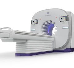
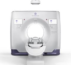
Sept. 30, 2024 — Siemens Healthineers recently announced the Food and Drug Administration’s premarket approval (PMA) for the tomosynthesis or three-dimensional breast imaging technology of the Mammomat B.brilliant.
This approval, which includes new 3D image acquisition and image reconstruction technology, adds to the system’s already cleared features for full-field digital mammography, or two-dimensional breast imaging; breast biopsy; and titanium contrast-enhanced mammography. Other features improve patient comfort, enhance user workflow, and improve ergonomics compared to its predecessor system, the Mammomat Revelation.
“The FDA premarket approval of the Mammomat B.brilliant mammography system represents a milestone in our ongoing commitment to women’s health,” said Niral Patel, vice president of X-ray Products at Siemens Healthineers North America. “This revolutionary system not only provides healthcare institutions with significantly improved diagnostic capabilities but also addresses the critical need for patient and technologist comfort in breast cancer screening.”
All Siemens Healthineers 3D mammography platforms use 50-degree wide-angle technology. This technology overcomes the issue of overlapping breast tissue that is a challenge with 2D imaging. It separates overlapping layers of breast tissue to help visualize lesions that would otherwise be obscured by providing up to 3.5 times the depth resolution (or tissue separation) of some other mammography systems.1
The Mammomat B.brilliant builds on this wide-angle approach with PlatinumTomo, a new 3D technology that enables a 50-degree acquisition in less than five seconds. PlatinumTomo reduces the blur inherent in 3D imaging and potentially assists the radiologist in making a more confident diagnosis. These benefits are possible due to the system’s fast detector and its new X-ray tube, which uses flying focal spot technology adapted for breast imaging from Siemens Healthineers computed tomography scanners.
Additionally, new UltraHD image reconstruction technology reduces metal artifacts, leverages for crisp visualization of calcifications, and offers customizable image settings. The system also provides a synthetic 2D image with no additional radiation exposure to the patient, reducing the radiologist’s need for full-field digital mammography images.
Several previously cleared features benefit the radiologic technologist. A prominent display monitor allows the technologist to clearly see patient information and work steps. The automated ComfortMove ergonomic feature enables the technologist to move the tube head independently of the image receptor, allowing for easy patient access. A laser guide permits accurate breast positioning. Together, these features can improve workflow.
An ergonomic hand grip and optimized, stationary face shield allow the patient to lean into the system during image acquisition for greater stability and the visualization of more posterior breast tissue. These features combine with a redesigned ambient light display, intelligent personalized breast compression, and rounded breast compression paddles to reduce patient discomfort, improve breast positioning, and enable more consistent image quality.
Additional information on the Mammomat B.brilliant can be found by clicking here.
1. Maldera, et al. (2016): Digital breast tomosynthesis: Dose and image quality assessment. Physica Medica, pp. 1-12.

 September 30, 2026