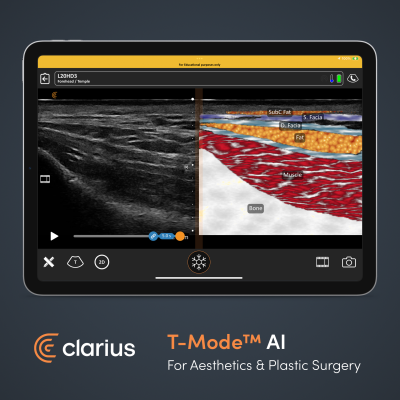
February 15, 2024 — Clarius Mobile Health, a global leader in wireless ultrasound solutions, announces T-Mode AI, a groundbreaking educational technology to help clinicians who are new to ultrasound advance their image interpretation skills using Clarius handheld scanners. Powered by artificial intelligence (AI), the new mode enhances a grayscale image using distinctive colors, patterns, and labels to teach clinicians how to instantly identify anatomical tissues and structures during an ultrasound exam.
"T-Mode is the beginning of a new era of ultrasound learning where AI acts as our "teacher" or "guide",” says Dr. Stefania Roberts, a phlebologist and experienced cosmetic physician practicing in Melbourne, Australia. “The learning curve to master ultrasound is steep, but with T-Mode, one can examine the temple region, lips, and cheek whereby fat, muscle, SMAS and bone are shown in different colors and then labelled so the user can confirm what the different layers in the face look like."
Wireless and pocket-sized, Clarius handheld ultrasound scanners deliver the high-definition imaging and performance of traditional ultrasound systems for a small fraction of the cost. They are the leading choice for plastic surgeons and aesthetics practitioners performing ultrasound-guided procedures to ensure patient safety.
Dr. Pat Pazmiño who has been using Clarius at his plastic surgery practice in Florida for 6 years believes T-Mode AI will enhance learning of fat grafting techniques: “Clarius continues their commitment to plastic surgery education with the introduction of T-Mode. This new interface allows surgeons to practice identifying the different subcutaneous layers and planning their fat grafting targets before they go to the OR.”
T-Mode AI is available now for Aesthetics and Plastic Surgery applications with Clarius wireless ultrasound scanners. Additional anatomical models supporting more medical specialties will be released over time. T-Mode AI is intended for educational and training purposes only. It is not intended for diagnostic use, interventional use, to guide injections, or for filler detection. Current users with Membership can access the new feature through the latest version of the Clarius App.
“The engineering team at Clarius has spent nearly a decade pushing the innovation barriers of ultrasound; we’ve removed wires, improved image quality and shrunk scanners to the size of an iPhone. And we’ve been using AI to make them easy to use. But I believe T-Mode is the most ground-breaking technology we’ve seen since the invention of B-Mode in the 70s and Color Doppler in the 80s because it truly makes ultrasound easy to learn for novice users,” says Clarius founder Laurent Pelissier. “Now we’re very excited to bring T-Mode to market to help more users unlock the power of ultrasound to deliver the best patient care.”
For more information: www.clarius.com

 June 15, 2026


