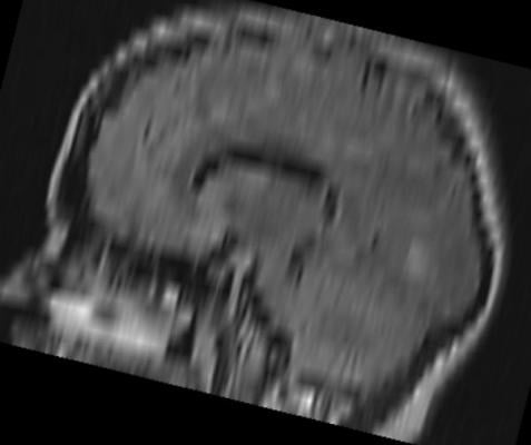
June 1, 2015 — Researchers from the Canada North Concussion Network in Manitoba examined neuroimaging studies obtained in children and adolescents with sports-related concussions (SRCs) and found that the images appeared normal in 78 percent of cases. Detailed findings of this study are reported and discussed in “Neuroimaging findings in pediatric sports-related concussion” by Michael J. Ellis, M.D., and colleagues, published online, ahead of print, in the Journal of Neurosurgery: Pediatrics.
Expert opinion among physicians specializing in sports-related concussion (SRC) holds that computed tomography (CT) scans and magnetic resonance images (MRIs) are not that useful in diagnosing SRCs. Until now, however, no study had been performed to verify this opinion.
The authors examined medical records and neuroimaging findings in 151 children and adolescents who had sustained SRCs during competitive sports activities such as hockey, soccer and baseball. The SRCs were all diagnosed and followed up by a single neurosurgeon specializing in concussion spectrum disorders at a multidisciplinary concussion program in the Pan Am Clinic in Winnipeg, Manitoba, Canada, between Sept. 1, 2013, and July 31, 2014. All cases were referrals from other physicians or athletic advisors.
Thirty-six patients (24 percent of 151 patients with diagnosed SRC) underwent neuroimaging. CT scans were obtained prior to referral in 23 patients and after consultation in one patient. Because CT scans were primarily obtained before referral and at a variety of facilities, the authors could not address the clinical indications for obtaining these CT scans. MRIs were obtained after referral to the concussion program in 15 patients and before referral in one patient. MRIs were ordered when patients displayed focal neurological deficits or symptoms lasting more than 1 or 2 months, and when there were abnormal findings on CT scans. In four patients both CT scans and MRIs were obtained. Abnormal CT and/or MRI findings were only found in eight (22 percent) of the 36 patients who underwent neuroimaging.
Abnormal findings were found on CT scans in five patients (skull fracture in two patients and suspected intracranial hemorrhage, arachnoid cyst, and suspected hemorrhage into an arachnoid cyst in one patient each) and on MRIs in four patients (intraparenchymal hemorrhage and sylvian fissure arachnoid hemorrhage; nonhemorrhagic contusion; demyelinating disease; and posterior fossa arachnoid cyst, cerebellar volume loss and nonspecific changes in white matter).
The authors discuss the common use of CT scans and the risks that excessive exposure to radiation may pose for children and adolescents. The fact that CT scans yield no signs of traumatic injury to structures of the brain in most cases of SRC leads the authors to suggest that use of CT should be limited to the emergency room setting in evaluating acutely injured patients in whom clinical signs or symptoms suggest the possibility of skull fracture or intracranial hemorrhage.
MRI findings of traumatic structural damage in conjunction with clinical signs and symptoms in two children led the neurosurgeon to advise against further participation in contact sports. Descriptions of these two cases are included, providing the reader with the array of clinical and neuroimaging factors that led to the physician’s advice.
Although neuroimaging does not reveal pertinent findings in most cases of SRC, the authors stress “the need to consider [magnetic resonance imaging] in pediatric patients with focal neurological deficits, worrisome symptoms, or abnormal of inconclusive CT findings,” adding that magnetic resonance imaging “should also be considered in pediatric patients with persistent symptoms for which the definition is unclear.”
When asked to describe the importance of the study, Ellis responded, “This study provides preliminary evidence that neuroimaging findings are normal in a significant proportion of pediatric sports-related concussion patients, but not every patient, and that neuroimaging can be helpful in informing clinical and return-to-play decision making in selected patients presenting with neurological symptoms following sports-related head injury.
“Methodologically, this study does not tell us which patients are more likely to demonstrate traumatic abnormalities on clinical neuroimaging, including magnetic resonance imaging. This question will be addressed by a prospective clinical study that is currently underway at our institution.”
For more information: www.thejns.org

 October 07, 2026