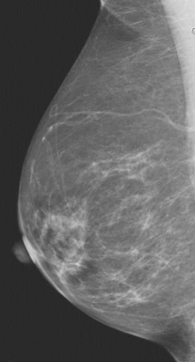
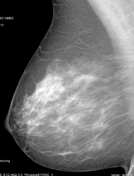
Fatty breast tissue.
December 3, 2013 — Automated breast density measurement is predictive of breast cancer risk in younger women, and that risk may be related to the rate at which breast density changes in some women as they age, according to research being presented at the Radiological Society of North America Annual Meeting (RSNA 2013).
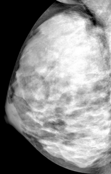
Breast density is already known to be a strong and independent risk factor for breast cancer. The American Cancer Society (ACS) considers women with extremely dense breasts to be at moderately increased risk of cancer and recommends they talk with their doctors about adding magnetic resonance imaging (MRI) screening to their yearly mammograms.
"Women under age 50 are most at risk from density-associated breast cancer, and breast cancer in younger women is frequently of a more aggressive type, with larger tumors and a higher risk of recurrence," Nicholas Perry, M.B.B.S., FRCS, FRCR, director, London Breast Institute, London, United Kingdom and the study’s senior author.
For the new study, Perry and colleagues compared breast density and cancer risk between younger and older women and analyzed how the risk relates to changes in breast density over time. The study group included 282 breast cancer cases and 317 healthy control participants who underwent full-field digital mammography, with breast density measured separately using an automated volumetric system.
"In general, we refer to breast density as being determined by mammographic appearance, and that has, by and large, in the past been done by visual estimation by the radiologist — in other words, subjective and qualitative," said Perry. "The automated system we used in the study is an algorithm that can be automatically and easily applied to a digital mammogram, which allows an objective and, therefore, quantitative density measurement that is reproducible."
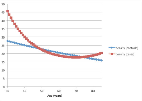
Breast cancer patients showed higher mammographic density than healthy participants up to the age of 50. The healthy controls demonstrated a significant decline in density with age following a linear pattern, while there was considerably more variability in density regression among the breast cancer patients.
"The results are interesting, because there would appear to be some form of different biological density mechanism for normal breasts compared to breasts with cancer, and this appears to be most obvious for younger women," said Perry. "This is not likely to diminish the current ACS guidelines in any way, but it might add a new facet regarding the possibility of an early mammogram to establish an obvious risk factor, which may then lead to enhanced screening for those women with the densest breasts."
For instance, some women might undergo a modified exposure exam at age 35 to establish breast density levels, Perry noted. Those with denser breast tissue could then be followed more closely with mammography and additional imaging like MRI or ultrasound for earlier cancer detection and treatment.
"It has been estimated that about 40 percent of life years lost to breast cancer are from women under 50 diagnosed outside of screening programs," said Perry. "In my practice, which is largely composed of urban professional women, 40 percent of cancers year to year are diagnosed in women under 50 and 10 percent in women younger than 40."
Co-authors are Katja Pinker-Domenig, M.D., Kefah Mokbel, M.B.B.S., FRCS, Sue Milner, B.Sc., and Stephen Duffy, M.Sc.
For more information: www.rsna.org
 October 19, 2026