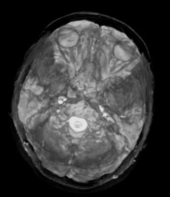
August 17, 2012 — Researchers at the Mayo Clinic say a common condition called leukoaraiosis, made up of tiny areas in the brain that have been deprived of oxygen and appear as bright white dots on magnetic resonance imaging (MRI) scans, is not a harmless part of the aging process, but rather a disease that alters brain function in the elderly. Results of their study are published online in the journal Radiology.
“There has been a lot of controversy over these commonly identified abnormalities on MRI scans and their clinical impact,” said Kirk M. Welker, M.D., assistant professor of radiology in the College of Medicine at Mayo Clinic in Rochester, Minn. “In the past, leukoaraiosis has been considered a benign part of the aging process, like gray hair and wrinkles.”
Leukoaraiosis, also called small vessel ischemia and often referred to as unidentified bright objects or “UBOs” on brain scans, is a condition in which diseased blood vessels lead to small areas of damage in the white matter of the brain. The lesions are common in the brains of people over the age of 60, although the amount of disease varies among individuals. “We know that aging is a risk factor for leukoaraiosis, and we suspect that high blood pressure may also play a role,” Welker said.
In the study Welker’s team performed functional MRI (fMRI) scans, a special type of MRI that measures metabolic changes in an active part of the brain, on cognitively normal elderly participants recruited from the Mayo Clinic Study of Aging between 2006 and 2010. In 18 participants, the amount of leukoaraiosis was a moderate 25 mL, and in 18 age-matched control participants, the amount of disease was less than five mL.
The patients were imaged in an MRI scanner as they performed a semantic decision task by identifying word pairs and a visual perception task that involved differentiating straight from diagonal lines. Although both groups performed the tasks with similar success, the fMRI scans revealed different brain activation patterns between the two groups. Compared to members of the control group, patients with moderate levels of leukoaraiosis had atypical activation patterns, including decreased activation in areas of the brain involved in language processing during the semantic decision task and increased activation in the visual-spatial areas of the brain during the visual perception task.
“Different systems of the brain respond differently to disease,” Welker said. “White matter damage affects connections within the brain’s language network, which leads to an overall reduction in network activity.” He pointed out that identifying leukoaraiosis in the brain is important, both for individual patients undergoing brain mapping for surgery or other treatments and for research studies. For improved neurological health, he said, efforts should be taken to prevent leukoaraiosis from occurring.
“Our results add to a growing body of evidence that this is a disease we need to pay attention to,” he said. “Leukoaraiosis is not a benign manifestation of aging but an important pathologic condition that alters brain function.”
For more information: www.radiologyinfo.org, www.rsna.org

 July 02, 2026


