June 8, 2010 - A multitracer molecular imaging technique using positron emission tomography (PET) provides detailed information about the physiological processes of cancerous tumors. The method also could eventually help radiation oncologists treat head and neck cancers with precision external-beam radiation therapy and improve the outcomes of therapy, according to research revealed at the Society of Nuclear Medicine’s 57th Annual Meeting, held June 6-8 in Salt Lake City.
“The research that we are conducting with Philips is extending the use of molecular imaging for radiotherapy planning, moving closer to more personalized treatment of hard-to-treat cancers based on the biology of each individual patient’s tumor,” said Kristi Hendrickson, Ph.D., lead author of the study and medical physicist at the University of Washington Medical Center, Seattle. “By modeling the data acquired from PET scans, we can potentially reduce damage to surrounding healthy tissue, as well as provide the ability to do ‘dose painting,’ delivering a highly customized form of radiation therapy for each patient.”
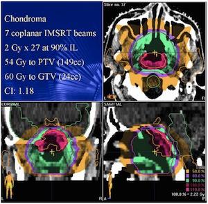
Cancers of the head and neck are notoriously difficult to treat, not only because of their proximity to sensitive anatomical structures, but also because of their tendency to recur. Researchers are working to find the best way to image these tumors in order to provide the most effective treatment. There are several forms of radiation therapy currently available. An approach called intensity modulated radiation therapy (IMRT) is a sophisticated technique that is used to maximize dose delivery to tumors while sparing adjacent normal tissues such as the salivary glands. This therapy uses an external beam of radiation that is sculpted into the shape and volume of the individual tumor. There are limiting factors involved in IMRT when applied to certain cancers. The presence of hypoxia, or oxygen depletion, can have a negative impact on therapy by leading to tumor resistance. By using biological information about the tumor gleaned from multiple PET imaging agents, clinicians can model both tumor anatomy and physiology, drawing closer to eradicating the cancer with a very high dose of radiation that is tailored to each tumor.
In this study, a patient with an advanced case of head and neck cancer showing hypoxia within the tumor was scanned using PET and two imaging agents, 18F-FDG, which measures glucose metabolism, and 18F-FMISO, which helps image and quantify hypoxia. Functional imaging with PET was combined with anatomical imaging from X-ray computed tomography (CT), and post-scanning analysis was used to model tumor physiology, using a Philips-developed pharmacokinetic modeling software. A high-dose IMRT plan was designed based on this information, which showed that treatment planning with molecular imaging is possible. This work may pave the way for more effective and individualized treatment for head and neck cancer patients. Further clinical trials evaluating patients treated with these approaches are needed to provide supporting evidence of the effectiveness of this and similar methods of IMRT treatment planning.
Reference: Scientific Paper 8: K.R. Hendrickson, U. Parvathaneni, J. Liao, Rad Oncology, University of Washington, Seattle, Wash.; M. Narayanan, Philips Research North America, Briarcliff Manor, NY; J.-C. Georgi, Philips Research Europe, Aachen, Germany; J.G. Rajendran, Radiology, U Washington, Seattle, Wash.; “Evaluating the feasibility of IMRT planning for head and neck cancer using dynamic F-FDG and F-FMISO,” SNM’s 57th Annual Meeting, June 5–9, 2010, Salt Lake City.
For more information: www.snm.org

 June 12, 2026


