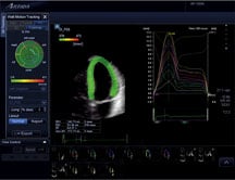
Normal dyssynchrony is demonstrated using Toshibas 3D wall motion tracking for the Aplio Artida ultrasound system. The left ventricle is color coded all green showing timing is in sync, while dyssynchrony would be color coded red.
Since the introduction of cardiac ultrasound, investigators have sought to quantify cardiac function in three dimensions. The recent development of a new technology called 3D wall motion tracking (3DWMT), which displays quantitative echo data from 3D ultrasound data sets, has tremendous potential to more accurately evaluate ventricular function in a wide variety of cardiovascular diseases.
At University Hospitals Case Medical Center in Cleveland, Ohio, we recently implemented 3DWMT in our clinical studies; we anticipate that removing subjectivity, minimizing interpretative variability and comprehensively and efficiently measuring strain, 3DWMT will revolutionize cardiac ultrasound diagnosis.
Benefits Over 2D Strain Imaging
The echocardiographic evaluation of regional myocardial function relies largely on the visual detection of endocardial wall motion abnormalities. However, this technique is subjective and demands complete visualization of the endocardium, and wall motion and thickening are subject to changes in cardiac loading and heart rate. Measurement of myocardial deformation (strain and strain rate) using 2-dimensional echocardiography has the potential to overcome these limitations. Because of 3D cardiac geometry and complex myofiber arrangements, there are multiple components or directions of strain (circumferential, longitudinal, and radial) that may be normal (i.e. orthogonal or perpendicular) or shear (i.e. torsional). While 2D strain (speckle tracking) and strain rate (tissue Doppler) imaging overcomes much of the subjectivity and variability inherent in assessing endocardial motion, these methods fail to address these complexities of cardiac geometry and motion. Not surprisingly, neglecting 3D wall motion may miss critical information and reduce diagnostic accuracy.
However, imaging a moving 3D structure, from a tomographic 2D plane creates obvious challenges. 3DWMT provides the physician and sonographer 3D depictions of the heart and graphical and parametric displays of myocardial deformation over time, all captured within a single cardiac cycle (4D mode) or from four stitched subvolumes (full 4D mode) and thus has the potential to meet these challenges. For example, using 2D acquisition methods there is the possibility of introducing error to strain measurements when the heart swings out of the imaging plane. When capturing the entire heart with 3DWMT, this issue is eliminated. An important advantage of 3DWMT is that the time for acquisition and analysis of longitudinal, circumferential, and radial strains is reduced. In addition, the effects of twisting (and torsion) motion can be quantified.
3DWMT for Ischemic Heart Disease
Based on these considerations, 3DWMT affords the potential for a more robust evaluation of cardiac function than does any 2D echocardiographic technique. Acquiring quantitative data in one heart beat eliminates the need to piece (or stitch) the data together spatially and temporally from separate beats and therefore may improve diagnostic accuracy. Owing to its sensitivity, strain analysis detects early, subclinical disease; as a result it has important potential applications in coronary artery disease, hypertensive heart disease, and cardiomyopathy.
One of the most important clinical applications of 3DWT is the assessment of ischemic heart disease. Ventricular volumes, ejection fraction and regional wall motion is rapidly assessed. Unique parameters of global ventricular systolic and diastolic function include rotation, twist and torsion. Global and regional vectors of strain and displacement in the radial, longitudinal and circumferential direction are simultaneously acquired from a single apical transducer position.
A major advantage of strain and strain rate imaging in wall motion analysis is the reduction of tethering and translational effects. 3DWMT
potentially identifies more accurately than 2D tethering of dysfunctional from normal myocardial segments, and abnormal wall motion due to cardiac translation. This is possible because the true 3D motion is contained within the ultrasound dataset, which eliminates the effects of through-plane motion that may occur with 2D imaging.
While stress echo is a proven clinical tool, the interpretation of segmental wall motion requires experience and expertise and in this setting is largely subjective. Strain and strain rate imaging have each shown promise as an adjunct to stress echocardiography; infarct-involved segments, visually normokinetic segments supplied by stenotic arteries, and regional ischemia are reliably detected. Strain measures may also be useful for the determination of myocardial viability, as stunned myocardium displays reduced systolic strain rates that improve with dobutamine infusion. Since 3DWMT provides an objective method to detect and quantify wall motion, the application potentially improves reproducibility when used in combination with stress echo. Detailed and quantifiable wall motion parameters may add to the accuracy of stress testing so the right patients are sent on for further testing.
3DWMT Diagnosis for Other Cardiovascular Disease
3DWMT is not only useful in the comprehensive assessment of myocardial deformation, but can potentially determine timing of deformation more accurately than 2D methods. Dyssynchrony of systolic strain as determined by 3DWMT may be valuable in the screening, selection and follow-up of patients who are candidates for cardiac resynchronization therapy (CRT). Fast acquisition and comprehensive information of all segments of the LV, facilitates the use of this method for patient selection and optimization of CRT devices immediately after implantation.
Since strains and strain rates are homogenously distributed across the myocardium, the detection of even subtle changes suggests myocardial dysfunction. Thus, 3DWMT has an important potential role in the diagnosis and management of virtually any disease that affects the myocardium. For example, anthracyclines are highly effective chemotherapeutic agents for breast cancer but their use is restricted by the frequent development of LV dysfunction and heart failure. Standard surveillance using the LV ejection fraction is often ineffective because the onset of clinical damage occurs late and the ejection fraction is load-dependent and fails to identify subclinical LV dysfunction. The ability of strain and strain rate imaging to efficiently monitor therapy, define the patient at risk, and predict later development of LV dysfunction and heart failure is exciting and promising. The ability for 3DWMT to give a more accurate representation of heart function may help physicians better make treatment decisions.
Although systolic strain and strain rates have received the greatest attention, changes in diastolic strain measures may also portend cardiac disease. For example, patients with restrictive cardiomyopathy have lower early diastolic strain rates than those with constrictive pericarditis. Strain rates measured in early diastole may also be able to differentiate hypertrophic cardiomyopathy and hypertensive left ventricular hypertrophy from physiologic hypertrophy in athletes.
Workflow and Patient Care
3DWMT has the potential ability to improve patient care and reduce health care costs by identifying heart disease more accurately and at an earlier stage. Studies have shown that 3DWMT is a simple and reproducible technique, and that compared to 2D speckle tracking, it more quickly and completely analyzes myocardial deformation. Coupled with rapid, semi-automated volume, LV ejection fraction and mass determinations, 3DWMT can increase the efficiency and productivity of the echo lab. As noted above, 3DWMT has the ability to detect subclinical disease and visualize small reductions in strain or strain rate as well as view subtle abnormalities in ventricular volume and function. Earlier diagnoses will also save valuable health care resources as disease can be treated in its earlier stages.
Complete analysis with 3D wall motion tracking can be completed in a few minutes and the method can be performed either on-line directly on the ultrasound system or off-line using a dedicated workstation. The acquired data is stored in a raw data format which allows off-line analysis with the same accuracy as the on-line procedure. Future parametric techniques can be retrospectively applied to stored datasets. We are performing our 3DWMT analysis on such a workstation housed in the echo reading area and are storing a Dicom clip of the analysis on our image analysis system.
The introduction of 3D wall motion tracking marks the beginning of a new era in cardiac ultrasound. Utilizing state-of-the-art ultrasound, cardiac function in full 3D information is now available and can be performed during the examination. 3D wall motion tracking provides clinicians with a more robust method to gain global and regional cardiac function information in everyday practice, including new and exciting parameters such as twist and torsion.

 July 13, 2026