September 1, 2009 - Evaluation of results of a multicenter study indicates that quantitative evaluation of the progression of volume of extracranial carotid vessel walls is feasible with 1.5T magnetic resonance (MR) imaging, despite limitations due to patient motion or habitus.
In the study, Atherosclerotic Plaque Progression in Carotid Arteries: Monitoring with High-Spatial-Resolution MR Imaging — Multicenter Trial, researchers set out to estimate the annualized rate of progression of vessel-wall volume in the carotid arteries in 160 patients by using MR imaging and to establish the fraction of studies that have acceptable image quality.
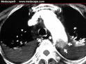
One hundred sixty patients with greater than 50 percent narrowing of the diameter of the carotid artery were recruited at six centers for prospective imaging of the carotid arteries at baseline and one year later by using high-spatial-resolution, 1.5T MR imaging. Quantitative changes in atheroma volume were measured on unenhanced T1-weighted images. A multiple linear regression analysis was used to correlate progression with several clinical factors, including statin therapy.
All 160 patients completed both baseline and follow-up studies. Of these studies, 67.5 percent were deemed to have image quality that was acceptable for quantitative analysis. The causes of rejection were motion (46 percent), deep location of the carotid artery (22 percent), low bifurcation of the carotid artery (13 percent), and “other” (19 percent). The mean annual change in vessel-wall volume was 2.31 percent. At one-year follow-up, vessel-wall volumes in patients not receiving statin therapy had increased faster compared with those in patients receiving statin therapy: 7.87 percent, plus/minus 13.58 percent vs 1.14 percent, plus/minus 9.9 percent, respectively.
Researchers found that in patients who had preexisting carotid disease, the rate of increase in vessel-wall volume was slower in patients receiving statin therapy.
Reference: Boussel Lo?c, MD, PhD., et al. Atherosclerotic Plaque Progression in Carotid Arteries: Monitoring with High-Spatial-Resolution MR Imaging—Multicenter Trial. Radiology: Volume 252: Number 3—September 2009. radiology.rsnajnls.org.
For more information: www.rsna.org

 July 10, 2026


