\"There will be...many advancements in PET, CT and MR technology, but the world will be changed by the molecules.
The field of molecular imaging continues to grow. GE Healthcare has already invested $160 million in the development of molecular imaging technology. Siemens Medical Solutions created a Molecular Imaging Division after its acquisition of CTI Molecular Imaging with the goal to further pursue development of hybrid imaging, preclinical systems and new biomarkers. Philips Medical Systems continues its commitment to PET and SPECT development, while at the same time has structured agreements with pharmaceutical companies, KEREOS and Theseus, to collaboratively establish camera design requirements to effectively image with specific molecular agents.
Pharmaceutical giants like Eli Lilly, GlaxoSmithKline, Novartis and Bristol-Myers Squibb are integrating molecular imaging into drug development. A new era of industry collaboration will continue to evolve as pharmaceutical and medical device companies work together on the development of molecular diagnostic tools.
One near-term goal of molecular medicine is to achieve personalized healthcare, where clinicians tailor drug therapies to individuals, and to bridge the gap between development and diagnosis of disease. Long-term, the goal is to “treat” the patient before a diseased or mutated cell divides and replicates – true predictive and preventive medicine.
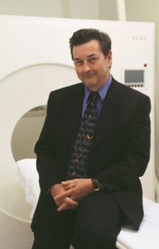
The significant advances in molecular medicine are outlined in Imaging Technology News’ exclusive interview with Michael Phelps, Ph.D., who is the chair of the Department of Molecular and Medical Pharmacology, UCLA; the director of Crump Institute for Molecular Imaging; associate director of UCLA-DOE Laboratory of Structural Biology and Molecular Medicine; chief of the Division of Nuclear Medicine, UCLA; and a Norton Simon professor and chairman.
Imaging Technology News: How do you define and view this expanding field of molecular imaging?
Dr. Michael Phelps: One way to look at molecular imaging is to look at the creation of it as a playing field. As you look on the playing field, you see all the positions on that field as a molecular something. So there is molecular technology, molecular biology, molecular diagnostics, molecular genetics, molecular therapeutics and molecular medicine – all of the world has gone molecular. Molecular imaging could be the partner to examine the molecular mechanisms of the biology of disease.
The focus and direction for molecular imaging is molecular therapeutics. Molecular therapeutics will become more defined about the direction of molecular imaging. If you look today, there are a number of things occurring that have an enormous impact on the years that lie ahead. First of all, the greatest science that will be done in the next 50 years is not high energy physics, but is biology and medicine. All the anatomical, physical and biological medical sciences come together to take on fundamental loss principals that govern biology, normal biology and its transformation into biology of disease.
ITN: There is much discussion on the “merger of biology and imaging.” Where do we go from here?
DMP: Biopharma has been growing tremendously. It is amazing that two years ago, it took 11 Genentech to equal one Merck. Today, it takes one Genentech to equal one Merck. There are about 1,500 biotech companies that are growing. But there are certain things that we need for molecular therapeutics to succeed. One of them is molecular diagnostics. With molecular diagnostics, either in vitro in the blood or in imaging, there are three things they want: [to know] by a direct measurement that the disease is present; [to get] information that tells me what stage it is in; and give feedback when I give a drug to the patient, a direct measurement of the impact that the drug has had on the molecular nature of the disease.
Every pharmaceutical company has a molecular imaging program or they have relationships with academic ones. To lead in those technologies and to sell those drugs in industry, the companies want to see in the image whether or not this patient has the drug part, the protein, particularly at a critical point. This is answered easily and routinely by imaging the target and measuring the biological process of that disease.
For example, every tumor does something strange when it goes through its transformations. It changes the way glucose is metabolized and does it very inefficiently. It doesn’t use the high energy capability that glucose can provide; it uses the first few steps of it, which is very inefficient. They design glycolosis so they can beat every cell in your body.
That’s a process infinitely led to by tumors, and that is why PET is so accurate in staging. There are other targets as well. There are molecules developed to image targets and separate patients by the drug charter. Another drug company does not know whether you have the drug charter and when you take your drug whether it hits that drug target. They don’t know if it requires 50 to 80 percent of that drug target to induce the pharmacologic effect, but they speculate with many years of research analysis.
There is another factor as well. Seventy percent of cancer patients are getting exposed to a drug with no benefits, but they incur the risk. We can’t afford that cost of the pharmaceutical process. Everyone has got to get their act together – the FDA, research programs, companies, etc. The goal is decreasing cost and increasing quality. The key to all of that is molecular diagnostics. What is the nature and stage of this disease, hence biology, and how does it respond to this drug? Everything goes back to molecular diagnostics. One portion in that is to develop these new diagnostics that take a drop of blood and examine protein signatures of the disease.
Another one is molecular imaging, going into the patient and finding an image to determine whether a certain protein target is there. The way the cells are reprogrammed to have disease is that the genome makes mistakes or has genetic errors. The mutated proteins wire the cell incorrectly so it functions differently. There are key proteins that allow these cells to shut off.
ITN: Where does nanotechnology fit into molecular
medicine?
DMP: Everything is valued through circuits. There is a new electronic system called molecular electrons or nanotechnology, where circuits are reprogrammed at the molecular scale. There is the Alliance for Nanosystems Biology, co-founded by Lee Hood, who did more than any single man in the world with the creation of the genomics era; Jim Heath, the king of nanotechnology; and myself. We put together the alliance and then we brought together a diverse group of people. The key to this is the system knowledge.
In front of you is a little chip about 2 cm that looks like an integrated circuit, but is not. It has the same sort of pattern, but it is in fluid space and there are channels, not wires, and all kinds of operations that can be performed in that chip. The size of the channels is about 30 to 40 microns. It has analysis aids, nanotech tools, chemistry labs to build and modify molecules, and it has a library to store molecules. We take that chip, hook it up to a PC and play the games that are biology – the games of disease and molecular therapeutics. How do we understand how to fix these things? If we go into resistance biology, you have to build integrated circuit technology, molecular electronics, to study this system biology. That is how you put it together. Remember we are 73 percent water, and then we have a bunch of cell cultures, and the cells are organized into certain cultures. Then we have a communications system, a vascular system that links it together. Basically we are cells and water connected on an integrated circuit that has tools to watch the DNA expression of instructions and formulation into protein circuits.
There is a new world called molecular targeted therapies, and in these therapies we manage the biology of the disease to improve and cure patients. If cells are saying things they should not be saying, for example, endless replication, we are going to clamp down on that cell.
ITN: How will this new world of molecular
targeted therapies work?
DMP: We’ve seen an example of two patients with enormous tumor growth diagnosed with pathological exams. They had the same disease and were both treated with the same drug. One of them had an unbelievable collapse of the tumors. In the other one, the tumor continued to grow. How can that be? We cannot predict the therapeutic response of mutation today because we don’t look at the right information. We have to look at the molecular basis of systems biology. With a CAT scan you give a test dose. If the patient responds, you continue; if the patient doesn’t respond, then you stop.
Take the example of a woman who has recurrent breast cancer; when we took out the breast tumor, we looked at the estrogen receptors and identified that she had high estrogen receptors. So we believe her cancer had metastasized. The primary tumor had high estrogen receptors, so we gave her hormone therapy. There were two images of this woman that we were looking at. One was glucose metabolism and another was fluroestrodial, the hormone that assesses the status of her estrogen receptors. Her metasis had high estrogen receptors, so we gave her hormone therapy. We then looked at two PET images, saw her metastasis, did the estrogen receptor study and found she had no estrogen receptors. We could give her hormone therapy, but it was not going to work. So what was going on here? Her primary tumor from which we made this decision did have high estrogen receptors, but her metastasis did not, which we saw in the PET images. How could it be? Metasis is not the same as a primary tumor, rather it moves on and gets worse. If we detected all cancers when they were primary cancers and they were in incapsulate, we could cure 98 percent of cancer right now. The majority of the time the cells are metasis that move on and get more malignant. So this case is an excellent example of what we learned by using PET before treating the patient.
ITN: What are you and your colleagues at UCLA
producing and how will this impact future clinical utility?
DMP: We have a unique culture [at UCLA]. As chairman of molecular medical pharmacology, we are using nuclear medicine in pharmacology and coming up with molecular diagnostics and therapeutics. These must be brought together. We have the Crump Institute which focuses on molecular imaging. We also have an institute called the Institute for Molecular Medicine. We bring together nanotech, systems biology and molecular imaging to create technologies and approaches. One one floor at the Institute for Molecular Medicine are clinical scientists conducting science that none of them can do on their own. They have to come together and work on molecular therapeutics and diagnostics in patients. They live a two-science concept, which is unusual. We are going to bring the basic and the clinical material into a single setting to fundamentally discover the therapeutic targets in the materials and the patients.
Then we have another unusual thing down at the L.A. tech centers. It is off campus, and half of it is nonprofit, the other half is for profit through Siemens [Medical Solutions]. Imagine if we developed the greatest drug to fight breast cancer and it stayed here in UCLA. It wouldn’t help anyone. So we have to get it out to the public through the industry.
The existing university world cannot deal with what’s going on today. The universities are organized in the old Germanic concept of chemistry, physics and biology. Companies are structured the same as well. They are not trying to solve the problem together. Our vision for how to do science is to create a culture where multiple disciplines live together and we have one simple rule: There are no rules. We are guided by trying to solve problems that impact the cure of patients. We are all passionately committed to this.
Take a scanner on your left, molecules on your right and in the middle of that are patients. There will be improvements and many advances in PET, CT and MR technology, but the world will be changed by the molecules. Everyone involved in medicine better wake up and realize the defying factors are in molecules. You have your mathematicians, engineers, chemists, immunologists, physicians – they all speak a different language. You bring them to a point where you say look, we must all become good teachers, and we must create a science that doesn’t exist today. Let’s create technologists to fulfill that. We can’t be instruments and molecules and we can’t be chemists and biologists. We can’t afford to have individual departments, both financially and in terms of making real progress.
You can keep improving the instruments, but you’ll change the world by improving molecules.


 February 06, 2026


