March 31, 2011 – Accurately navigating and interpreting cardiovascular anatomy for precise device deployment is a challenge to physicians during complex interventional procedures. In order to help solve that problem, the Infinix-I X-ray system, from Toshiba, has been upgraded.
Upgrades include Volume Navigation 3D roadmapping for all new systems, a 12-inch x 12-inch flat panel detector on ceiling mounted C-arms and the Maquet Magnus OR table availability for the Infinix VC-i and CC-i systems.
“Developing industry-leading technology that improves diagnostic accuracy, workflow and patient outcomes provide physicians with greater confidence when deciphering and navigating complex cardiovascular structures,” explained Doug Ryan, vice president, Marketing and Strategic Development, Toshiba. “These upgrades to the Infinix-i line of cardiovascular X-ray systems enable physicians to perform complicated interventions more quickly and accurately, another example of Toshiba’s commitment to improving patient care.”
Now available on all Infinix-i systems, the company’s real-time Volume Navigation 3D roadmap displays the deployment of coils during intervention on a cerebral aneurysm with exceptional clarity and precision. It links the movements of the system components with the fusion 3-D and fluoroscopic display. As a result, despite changes in table and C-arm position, the 3-D overlay is automatically aligned with the fluoroscopic image with high accuracy. In addition, it provides 2-D and 3-D roadmap display modes and allows physicians to fine-tune images with manual controls for device enhancement, further assisting physicians when making difficult decisions during advanced procedures. The feature is particularly helpful for procedures on intricate vascular regions, such as the brain, uterus and abdomen.
The Infinix CC-i and VC-i ceiling mounted systems are now available with a 12-inch x 12-inch flat panel detector. This midsized panel allows for an improved field-of-view, provides twice as much anatomical coverage compared with smaller flat panel detectors and is optimal for performing a range of cardiovascular diagnostic and interventional procedures, including those on pediatric patients. The small profile of this midsize detector is well suited to the operating room environment.
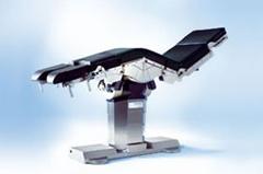
Additionally, the systems are now available to be combined with the Maquet Magnus table for enhanced use during hybrid OR procedures. As a result of this compatibility, rotational, low-contrast and 3-D angiography imaging can now be performed in rooms with the equipment. The system’s design is suitable for open surgical and catheter-based interventions, providing optimum patient access, a critical capability for hybrid exams.
For more information: www.maquet.com, www.medical.toshiba.com