Beth Whiteside, M.D., and her colleagues at Albany Medical Center worked with tomosynthesis during the research trial, and the center was the first facility in the country to announce plans to install breast tomosynthesis technology after it received FDA approval.
“Tomosynthesis really allows us to look through the breast, slice by slice, in a way that we haven’t been able to before,” explains Beth Whiteside, M.D., breast imaging specialist at Albany Medical Center and radiologist with Community Care Physicians. “It takes away that vexing issue of tissue overlap and allows us to look underneath. I think it is every radiologist’s dream.”
Located in Albany, New York, the Albany Medical Center began working with tomosynthesis during the research trial. Four days after the technology received U.S. Food & Drug Administration (FDA) approval, the Medical Center issued a press release announcing its intention to install breast tomosynthesis. “We jumped at the chance to implement it in practice,” states Whiteside. “We want to save lives, and we hope that tomosynthesis is going to help us do that.”
Increasing Confidence and Reducing the Recall Rate
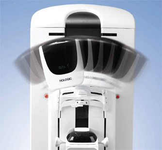
Albany Medical Center is using Hologic’s Selenia Dimensions digital tomosynthesis system for both screening and diagnostic mammograms. “Tomosynthesis has been very successful. It definitely increases our confidence and improves the visualization of cancerous lesions. Every radiologist has had a case where you see an area that looks very bright white and you’re wondering if there’s a mass underneath,” continues Whiteside. “Tomosynthesis really allows you to get at that. It’s almost instant satisfaction.”
An academic health sciences center, Albany Medical Center is dedicated to providing residents across eastern New York and western New England with access to the latest medical and technological innovations. The Center makes tomosynthesis available as a screening modality to all patients, based on availability of the system.
“I would love it if all of our machines were capable of doing breast tomosynthesis,” states Whiteside. Albany Medical Center also uses tomosynthesis as a diagnostic tool, in select cases, to guide the workup. “Tomosynthesis helps you determine with more confidence where the lesion is located. It gives you excellent characterization of many lesions,” she adds.
Tomosynthesis has helped the breast center staff find cancers they might not have seen with 2-D mammography. The technology is also helping to reduce the recall rate.
“One of the difficulties with regular screening mammography is tissue overlap,” explains Whiteside. “Tomosynthesis gives radiologists a lot more information and helps us deal with the issue of superimposed tissue. You can look through the tissue and make sure there’s nothing underneath. It gives us a lot more confidence as we see the cancerous lesions spring to life on the screen, and that has definitely reduced recall rates.”
A Seamless Transition for Patients and Radiologists
Patients experience very little difference between a 2-D and breast tomosynthesis mammogram. “Tomosynthesis is almost invisible to the patient,” adds Whiteside. “It’s much like getting a regular 2-D mammogram; tomosynthesis is no more inconvenient or uncomfortable for the patient. But the added advantage is all the extra information the radiologist gets. I think that’s particularly exciting for people who either have a personal history of breast cancer or know someone who has been touched by breast cancer.”
Some patients do ask about the radiation dose for the tomosynthesis exam. But Whiteside assures them that the dose is well within the guidelines the FDA sets for regular 2-D mammography.
For Whiteside, mammography is definitely the gold standard. “But one of the things that’s great about breast tomosynthesis is it is based on mammography, a technology that we’ve got a ton of information about already. That allows us to implement it into our practice in a seamless way,” she says. “I think that radiologists, as scientists, will want to see the data and know that large trials support the use of tomosynthesis. Certainly the initial evidence is very encouraging, and I think that you’ll see adoption of the technology go along with the data.”
Concludes Whiteside, “The first time you see a case of breast cancer that is very difficult, almost impossible to see on a regular 2-D mammogram pop out on the tomosynthesis images, you become an instant believer.”
The views and opinions expressed herein are those of Dr. Whiteside and her colleagues at Albany Medical Center and are not necessarily those of Hologic. This information is intended for medical professionals in the U.S. and other markets and is not intended as a product solicitation or promotion where such activities are prohibited. Because Hologic materials are distributed through websites, eBroadcasts and tradeshows, it is not always possible to control where such materials appear. For specific information on what products are available for sale in a particular country, please contact your local Hologic representative or write to [email protected].
Hologic, Dimensions and Selenia are trademarks and/or registered trademarks of Hologic and/or its subsidiaries in the U.S. and/or other countries.