January 29, 2008 - The PEM/PET system, a new medical imager for detecting and guiding the biopsy of suspicious breast cancer lesions, is capable of spotting tumors that are half the size of the smallest ones detected by standard imaging systems, according to a new study.
The results of initial testing, designed and constructed by scientists at the Department of Energy's Thomas Jefferson National Accelerator Facility, West Virginia University School of Medicine and the University of Maryland School of Medicine will be published in the journal Physics in Medicine and Biology on Feb. 7.
Testing of the new imager was led by Ray Raylman, a professor of radiology and vice chair of Radiology Research at WVU and lead author on the study. Raylman's team imaged various radioactive sources to test the resolution of the system.
“We had good performance characteristics, with image resolution below two millimeters. In regular PET, the image resolution is over five millimeters, so we're quite a bit better than that,” Raylman said. In addition, the initial tests revealed that the PEM/PET system can complete an image and biopsy in about the same amount of time as a traditional biopsy.
“The ability of the device to do biopsy is probably one of its most unique characteristics. There are other breast imagers, but none that are built specifically to do biopsy as well as imaging,” Raylman said.
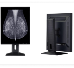
The system features components designed for imaging the unique contours of the breast. Known as positron emission mammography (PEM), this imaging capability enables users to attain high-resolution, three-dimensional PET images of the breast. The PEM/PET system images the breast with a movable array of two pairs of two flat detection heads.
If a suspected lesion is found, a single pair of heads is then used to guide a needle biopsy of the lesion; the biopsy is performed with a person-controlled robot arm. Raylman is the author of the concept and has a patent on this idea. The system is especially useful in imaging tumors in women who have indeterminate mammograms because of dense or fibroglandular breasts.
The Jefferson Lab Radiation Detector and Medical Imaging Group, with a group member now affiliated with the University of Maryland School of Medicine, developed the detector heads with the on-board electronics, the data acquisition readout and the image reconstruction software. The imaging device's gantry and the motion-control software were developed by West Virginia University researchers.
The next steps for the team include minor improvements in the detector systems and image reconstruction software and the addition of components for taking x-ray computed tomography (CT) scans. Initial clinical trials are planned after completion of system testing.
For More Information: www.jlab.org
 March 12, 2024