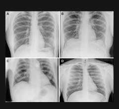

October 17, 2017 — At the 2017 Radiological Society of North America Annual Meeting (RSNA 2017), Konica Minolta will demonstrate how the KDR Advanced U-Arm (AU) System can help radiology practices improve workflow and patient satisfaction while providing impressive clinical images to help clinicians make better decisions sooner.
Advanced design innovations on the KDR AU optimize workflow, increase staff efficiency and improve outcomes to expedite the diagnostic process and elevate the patient experience. Innovations include:
- The system automatically swivels into position across a 135-degree range of motion and 35 inches of vertical movement, saving valuable time;
- The advanced detector and Ultra acquisition software work quickly in unison to enhance the acquired image; and
- Information is instantly retrieved, prompting the system to move to a predetermined position and source-to-image-receptor distance (SID) to save time when positioning the patient.
The versatile, compact design of Konica Minolta’s KDR Advanced U-Arm System makes it ideal for facilities that need to provide superior-quality radiography services in relatively small spaces.
In addition, the company will introduce the 10 x 12-inch and 17 x 17-inch versions of the AeroDR HD digital radiography (DR) detector panel. The 10 x 12 compact size is ideal for meeting the specific needs of extremity imaging and neonatal intensive care unit (NICU) environments, and the 17 x 17 is designed to be mounted in a semi-permanent location for imaging larger anatomical areas, such as the chest and abdomen.
All AeroDR HD Panels offer the option to switch between high definition (HD) and high dynamic range (HDR) imaging. High definition uses 100μ to give more detail of small structures. Konica Minolta’s image processing enhances sharpness and contrast while keeping the natural granularity of the structures, like trabecular bone, even under magnification. High-definition is recommended for extremities, shoulders and other structures where magnification is preferred to improve image quality for diagnostic accuracy.
High dynamic range aggregates the data from 4 pixels for a wider range of grays, providing a smooth image that allows clinicians to detect subtle differences in soft tissue.
For more information: www.konicaminolta.com/medicalusa
See what products vendors plan to display at RSNA 2017 on ITN's RSNA FastPass microsite at www.rsnafastpass.com. Click on the top dropdown menu to choose products by modality.

 October 07, 2026