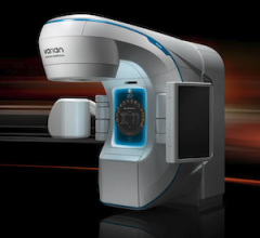
February 2, 2010 - Researchers found that while using Gamma Knife radiosurgery was well-tolerated as initial therapy in the management of patients with sarcomatous intracranial metastases, many patients end up developing progressive brain disease.
Researchers wanted to determine the indication and outcomes for Gamma Knife stereotactic radiosurgery (GKSRS) in the care of patients with intracranial sarcomatous metastases. To do so , they looked at data from 21 patients who underwent radiosurgery for 60 sarcomatous intracranial metastases (54 parenchymal and 6 dural-based) were studied. Nine patients had radiosurgery for solitary tumors and 12 for multiple tumors.
The primary pathology was metastatic leiomyosarcoma (4 patients), osteosarcoma (3 patients), soft-tissue sarcoma (5 patients), chondrosarcoma (2 patients), alveolar soft part sarcoma (2 patients), and rhabdomyosarcoma, Ewing’s sarcoma, liposarcoma, neurofibrosarcoma, and synovial sarcoma (1 patient each). Twenty patients received multimodality management for their primary tumor, and 1 patient had no evidence of systemic disease. The mean tumor volume was 6.2 cm3 (range, 0.07–40.9 cm3), and a median margin dose of 16 Gy was administered. Three patients had progressive intracranial disease despite fractionated whole-brain radiotherapy before SRS.
They found that a local tumor control rate of 88 percent was achieved, including patients receiving boost, up-front, and salvage SRS. New remote brain metastases developed in 7 patients (33 percent). The median survival after diagnosis of intracranial metastasis was 16 months, and the 1-year survival rate was 61 percent.
Although the therapy was well-tolerated in the management of patients with sarcomatous intracranial metastases, they found that many patients, including those who also received fractionated whole-brain radiotherapy, developed progressive new brain disease.
Reference: Flannery, T; Kano, K; Niranjan, A. Gamma Knife Radiosurgery as a Therapeutic Strategy for Intracranial Sarcomatous Metastases. Int J Radiat Oncol Biol Phys. 2010 Feb 1;76(2):513-519.
For more information: www.oncologystat.com

 October 23, 2026