Part of Sentara's comprehensive marketing campaign for 3D mammography included interactive community outreach initiatives.
When Sentara Healthcare decided to offer 3D mammography, also known as breast tomosynthesis, across its network of comprehensive breast centers in Virginia, it adopted the technology at 10 breast centers over 12 months. It was a successful introduction based on a carefully orchestrated implementation strategy. The technology would impact many aspects of the Sentara Cancer Network, requiring close collaboration and coordination with Sentara leadership, breast center staff, private radiologists and surgeons, as well as Sentara IT, finance, marketing and communications, hospital administrators, referring physicians and patients.
Sentara installed its first Hologic Selenia Dimensions 3D mammography system in January 2013, adding a second site in April. The remaining eight Virginia systems were installed through the end of that year. “It was a huge feat to coordinate the purchase of 3D mammography across Sentara, which is located in multiple regions of Virginia and now part of North Carolina,” stated Joanne M. Inman, MHA, FACHE, vice president operations, Sentara Virginia Beach General Hospital, who also serves as leader for breast cancer services.
The Sentara Cancer Network is among the nation’s first accredited cancer network programs through the American College of Surgeons and operates 20 breast centers in the Hampton Roads and Northern Virginia regions of the Commonwealth. In these regions, Sentara performs 120,000 mammograms annually, and was the first provider in Hampton Roads to offer 3D mammography.
“We diagnose about 1,600 new breast cancer cases a year,” stated Cynthia Allen, RN, Sentara vice president cancer services. “That’s 22 percent of all breast cancer cases diagnosed throughout Virginia.”
Collaboration is Key to Successful Implementation
The Sentara culture of readily adopting innovations and its organizational structure were key drivers to implementing cutting-edge technology across a geographically dispersed network. Within the Sentara Cancer Network, cancer specialists work as a multi-disciplinary team to provide comprehensive, innovative care from prevention through treatment. It was the Cancer Network’s Breast Program Leadership Committee that led the way in the evaluation, advocacy and adoption of 3D mammography. The committee includes surgeons, radiologists, pathologists, radiation oncologists, medical oncologists and navigators, all partnering with breast center leaders.
“As soon as the FDA approved tomosynthesis, a group of our radiologists brought it before the Breast Program Leadership Committee, and we recommended it to Sentara corporate leaders,” explained Kelley Allison, M.D, fellowship-trained mammographer and co-chair of the Sentara Breast Program Leadership Committee. “The ultimate catalyst was the strong support of our physician partners who advocated for the adoption of this technology based on early clinical findings,” added Allen.
Selecting Pilot Sites
Sentara leadership decided on a phased implementation to allow time to evaluate and model national results in its own facilities.
“For the first phase, we looked at the geographic areas with the highest incidence of breast cancer and the centers performing the greatest volume of mammograms,” explained Inman. “In addition, we considered locations with strong physician champions and aging existing mammography equipment. Another key decision prior to implementing 3D mammography was the best clinical application of the technology. After careful consideration, Sentara decided to offer 3D exams to all screening patients, with a patient co-pay.
Building a Case for Full-scale Implementation
Once the two pilot sites were up and running successfully, the Breast Program Leadership Committee went back to Sentara leadership for approval of additional sites, backed by real case histories, several months of data and the shared experiences of the other institutions that had already adopted the technology. “When we initially recommended 3D mammography, large-scale research studies weren’t available,” explained Allison. “But, by the time we were ready to add additional sites, research studies were showing the 3D technology could lower recall rates and improve cancer detection, especially with invasive breast cancers. Data from our pilot sites correlated with the national studies; we were finding more cancers with 3D. In fact, on day one we diagnosed a case that we would not have seen without 3D. Once we presented that data, we received support to implement 3D mammography across the system.”
“During the course of the conversation, Sentara leadership asked whether 3D would help increase volumes,” added Inman. But, most importantly, we looked at the clinical outcomes with 3D. The clinical efficacy was a major factor and most compelling in the decision to move forward with 3D to help further our mission of improving health every day.”
Managing Growth and Expectations
With approval of the additional sites, the Breast Program Leadership Committee turned to operationalizing its plan. This included developing IT infrastructure, providing training for physicians and staff, designing process changes and developing communications for the referring physician and patients.
“With only one Hologic 3D unit in each center, we’re considering additional units to meet the growing patient demand,” said Allison. “Not a day goes by that we’re not finding these small cancers for our patients; 3D makes our exams more accurate. We want to offer that option to even more women.”
With a single 3D system in each center, scheduling appointments initially posed challenges. The breast centers had to adapt their scheduling systems and encourage patients to ask for 3D exams when they make their appointment. If a patient chooses to convert to 3D at the time of their appointment, and other patients are already scheduled on the system, it significantly impacts the workflow. Offering 3D also required changes to the way the exam is coded, which requires coordination between billing and legal departments to implement the advance beneficiary notice (ABN).
“We made sure all breast center staff members were trained and well versed on exactly what to say about 3D mammography,” explained Allison. “We trained our front desk personnel, technologists, people in our file room — really anyone who could answer a telephone or may have to answer a question about 3D mammography.”
Today, patients are increasingly choosing the 3D exam. The two pilot centers are seeing a 40-50 percent conversion rate. “About half of the mammograms done at our facility today are 3D,” reported Allison.
To accommodate the size and number of 3D images, Sentara needed to upgrade the bandwidth and network speed, and increase data storage capabilities at the breast centers. “We involved our IT team in the process from the beginning,” stated Inman.
Educating a Community
Sentara helped inform key referring physicians with information packets containing FAQs, studies on 3D and a letter from a lead radiologist at the local breast center. Physician liaisons, breast center managers and radiologists also talked with key referring physicians.
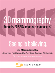
In addition, Sentara implemented a comprehensive marketing and communications campaign that involved a combination of earned and paid media in all major outlets. The plan also included engaging consumers in social media campaigns and innovative marketing efforts such as interactive digital billboards and displays to inform the community about the benefits of 3D mammography and the availability of the exam at Sentara breast centers.
Building on a Successful Implementation
In the end, the implementation of 3D mammography required a significant investment of time with affected people and teams across Sentara. Contributing to the system’s success was incorporating learnings from the two pilot sites to ease the next phases of adoption. Inman’s advice to other organizations preparing to implement the cutting-edge technology is be prepared to invest time and involve all of your stakeholders. “We successfully implemented 3D throughout our network in a relatively short period of time, because we involved and collaborated with our physicians, administrators and support teams. What we accomplished is really substantial,” concluded Allen. “We were able to implement 3D as quickly as we did because of the formal structure of the Breast Program Leadership Committee and the involvement and commitment of every member of the Sentara team.”
Case study supplied by Hologic Inc.
The views and opinions expressed herein are those of Sentara Healthcare and are not necessarily those of Hologic. This information is intended for medical professionals in the U.S. and other markets and is not intended as a product solicitation or promotion where such activities are prohibited. Because Hologic materials are distributed through websites, eBroadcasts and tradeshows, it is not always possible to control where such materials appear. For specific information on what products are available for sale in a particular country, please contact your local Hologic representative or write to [email protected].
Hologic, Dimensions and Selenia are trademarks and/or registered trademarks of Hologic and/or its subsidiaries in the U.S. and/or other countries.

 July 07, 2026


