Diagnostic Imaging Northwest Announces the First Extremity MRI Scanner in Northwest

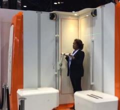
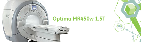
February 6, 2012 — Diagnostic Imaging Northwest (DINW) in Tacoma, Wash., announced the new extremity 1.5 Tesla magnetic resonance imaging (MRI) scanner that offers new comfort for patients.  The GE Optima is the first specialty high-field extremity MRI available in the Northwest. Designed to be more comfortable than a full-body MRI system, the GE Optima is intended for patients needing an MRI exam of extremities such as the elbow, wrist, hand, knee, ankle and foot.

The Optima features a high-strength 1.5T magnet exceeding those of a standard MRI system, and warranting image quality. The comfortable design ensures less moving around; resulting images are likely to be even more clear and consistent, securing a more confident diagnosis.

Unlike the awkward and uncomfortable positions sometimes required for extremity scanning in whole-body systems, patients can relax on a padded chair beside the scanner, reclining comfortably. The open design eliminates anxiety for patients who experience extreme claustrophobia with traditional closed-in tube MRIs.

Barbara Blankenship, medical director for DINW, said, "Our goal is to provide patients with the right MRI scanner for their needs. Comfort and quality imaging are our top priority. For some extremity exams done in a traditional full-body MRI system, the way the patient must be positioned is not always comfortable." Beyond comfort, the extremity MRI's open design reduces anxiety for those who have concerns about going into a full-body MRI system. Additionally, children can be joined by an adult in the exam room.

"With the GE Optima MRI, patients sit next to the system and only the targeted body part goes into the system. This helps make it easier for them to be as still as possible for the best exam achievable," Blankenship said. 


Related Content

News | PACS

April 28, 2020 – Konica Minolta Healthcare Americas released its Rede PACS, the company’s newest medical image ...

Time April 28, 2020
arrow
Videos | Orthopedic Imaging

This is a demo of the EOS orthopedic X-ray imaging system at the recent 2019 Radiological Society of North America (RSNA ...

Time January 03, 2020
arrow
News | Orthopedic Imaging

October 24, 2019 — EOS imaging announced the first patient cases performed with its hipEOS 3.0 surgical planning ...

Time October 24, 2019
arrow
News | Orthopedic Imaging

International medical imaging information technology (IT) and cybersecurity company Sectra is extending its pre-operative orthopedic planning solution for trauma cases with 3-D templates from one of the largest orthopedic companies, DePuy Synthes. Pre-operative planning for trauma cases using templating in combination with 3-D computed tomography (CT) images enables improved surgical outcomes.

Time October 17, 2019
arrow
News | Mobile C-Arms

Digital medical technology company Brainlab unveiled Loop-X, which it calls the first mobile intraoperative imaging robot, at the 2019 North American Spine Society annual meeting (NASS 2019), Sept. 25-28 in Chicago.

Time September 26, 2019
arrow
News | Orthopedic Imaging

September 5, 2019 — An ahead-of-print article published in the December issue of the American Journal of Roentgenology ...

Time September 05, 2019
arrow
News | Ultrasound Imaging

Konica Minolta Healthcare announced a new autologous biologics workshop in partnership with EmCyte Corp. The workshop is part of Konica Minolta Healthcare’s UGPro Solution, an initiative that provides complete solutions for ultrasound-guided procedures and therapies featuring the company’s Sonimage HS1 and Sonimage MX1 ultrasound systems, and including hands-on education and training.

Time June 12, 2019
arrow
Technology | Artificial Intelligence

Radiology artificial intelligence (AI) provider Aidoc announced the U.S. Food and Drug Administration (FDA) has cleared the world’s first AI solution for triage of cervical spine fractures. The latest 510(k) clearance is Aidoc’s third, coming just weeks after the FDA cleared Aidoc’s pulmonary embolism solution. By prioritizing suspected C-spine fracture cases in a radiologist’s worklist, Aidoc’s AI-powered triage automatically prioritizes critical cases, ensuring they are diagnosed by a radiologist in the most timely manner.

Time June 11, 2019
arrow
News | Ultrasound Imaging

May 29, 2019 — QT Ultrasound recently showcased its advanced ultrasound technology at the Acoustical Society of America ...

Time May 29, 2019
arrow
News | Ultrasound Imaging

The Hong Kong Polytechnic University (PolyU) announced the development of a palm-sized 3-D ultrasound imaging system for radiation-free scoliosis assessment, dubbed Scolioscan Air. The system can bring accurate, safe and cost-efficient mass screening to schools and anywhere in the community.

Time May 03, 2019
arrow
Subscribe Now