
X-ray Has Come a Long Way in 100 Years
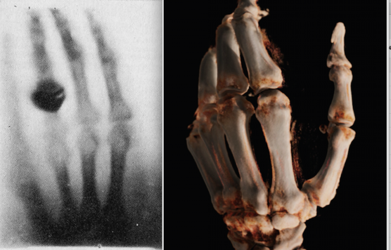
Left, the first X-ray ever made of Roentgen's wife's hand in 1895. Right, a cone-beam CT 3-D reconstruction of a hand in 2015 using a new robotic digital radiography (DR) X-ray system.
Being an avid student of history, I am always looking for parallels and comparisons in everything I see. For this reason, I was very struck by the latest X-ray technology displayed at the 2015 meeting of the Radiological Society of North America (RSNA) in December. Being in its 101st annual RSNA meeting, and the 120th anniversary of the discovery of X-rays, you would think that there is not much new in regards to X-ray technology. However, one of the images included in a Siemens press kit for their new robotic X-ray room technology, for me, brought the world of radiology full circle from its inception more than a century ago, to the bleeding edge of medical technology today.
Two weeks after Wilhelm Roentgen first discovered what he termed as X-rays in 1895 (he used the mathematical “X” to describe something unknown), he produced the first X-ray image of his wife’s hand. This image was the first medical imaging photo published in the first scientific article on medical imaging in December 1895. The breakthrough technology rapidly revolutionized medicine and earned Roentgen the first Nobel Prize in physics in 1901.
Being the symbol of the birth of radiology and modern medical imaging, this image of Roentgen’s wife’s hand was the first thing I thought of when I ran across an image of a cone-beam computed tomography (CT) 3-D reconstruction of a hand created by Siemens' new Multitom Rax robotic X-ray system. The comparison of hand X-rays now and then is a simple comparison of how far X-ray technology has advanced, from a fuzzy image of phalanges to a surgical, photo-quality view of the bone.
The Multitom Rax room installed system uses two robotic arms to precisely align the X-ray tube and detector panels in any position. It is designed to be an all-in-one X-ray room solution for conventional 2-D radiography, fluoroscopic exams, basic angiography applications and to create 3-D cone-beam CT images. The cone beam CT technology uses a series of X-rays shot in an arc around the patient to collect a volume of data, similar to a CT scanner collecting a volume of data through a series of scan slices. The computer can then post-process the cone beam dataset into 3-D image reconstructions.
Up until recently, dedicated X-ray systems were used for specific types of X-ray applications such as angiography, CT, digital radiography or fluoroscopy. This is likely the first X-ray system to be able to fulfill all of these imaging applications (at least on a basic level) using one platform. Cone beam CT created from a series of X-ray images previously found a niche in the cath lab, where newer C-arm systems can perform a rotational angiography spin around a patient and a 3-D image of the anatomy can be created tableside for use as a guidepost to landmark anatomy not visible on angiography alone.
Cone beam CT is used for advanced dental imaging and as onboard 3-D imaging on some radiation therapy treatment systems. It is now finding a new niche in orthopedic imaging as a less expensive, lower-dose and immediately available option, rather than separate X-ray and CT exams. Carestream adapted its cone beam technology commercialized for the dental market to a larger system aimed at the orthopedics market at a fraction of the cost of a CT scanner. The new system was displayed for the first time at RSNA 2015.
Watch a video on some of the most innovative new imaging technology at RSNA 2015.
Related Content

News | X-Ray
July 14, 2026 — A team of crew members aboard a commercial spaceflight acquired the first diagnostic X-rays during an ...
 July 14, 2026
News | Mammography
June 23, 2026 — Using artificial intelligence (AI), researchers found that image-based risk scores for breast cancer ...
June 24, 2026 
Feature | X-Ray | Kyle Hardner
Water-window X-rays allow researchers to visualize biological cells at high contrast without staining agents or other ...
June 23, 2026 
News | Radiology Imaging
June 15, 2026 — Lead Glass Pro, a supplier of radiation shielding products, has expanded its turnkey installation ...
June 18, 2026 
News | Digital Radiography (DR)
June 3, 2026 – Konica Minolta Healthcare Americas has announced a new version of AeroRemote Insights, the company’s ...
June 05, 2026 
News | Computed Tomography (CT)
April 2, 2026 — Nano-X Imaging Ltd. recently announced its U.S.-based subsidiary, Nanox Impact Inc., has signed a new ...
April 08, 2026 
News | Radiology Business
March 31, 2026 — Radon Medical Imaging, a medical imaging equipment maintenance and repair services company, has has ...
March 31, 2026 
News | FDA
March 24, 2026 — MARS Bioimaging, a New Zealand–headquartered medical device company, has received U.S. Food and Drug ...
March 25, 2026 
News | Pediatric Imaging
March 17, 2026 – OXOS Medical recently announced that its MC2 portable X-ray system is now cleared for pediatric imaging ...
March 23, 2026 
News | Radiology Imaging
March 23, 2026 — Samsung Medison hsa announced that its U.S. medical imaging businesses, previously operating as ...
March 23, 2026 © Copyright Wainscot Media. All Rights Reserved.
Subscribe Now