During the American College of Cardiology 2013 (ACC.13) annual meeting in March, vendors discussed several trends they are observing in the cardiac ultrasound market and displayed the latest echo advances.
Automation is making ultrasound systems easier to use, helping shorten exam times by reducing the number of steps needed to perform measurements or to slice images to achieve a specific view. Automation also has simplified
the quantification and measurements, which are completed on some systems in seconds with just a couple of clicks.
Examples of this include GE Healthcare’s automated workflows to simplify common echo image crops and measurements, and other functions. Instead of requiring five or more clicks to perform a crop, GE’s systems have reduced this number down to one or two clicks. The company also introduced Automated Function Imaging (AFI), a software tool that automates 2-D speckle tracking to measure the deformation (strain) of the myocardial wall in real time. It is available on several GE Vivid products. GE said AFI is a more sensitive method for assessment of left ventricular function than ejection fraction (EF).
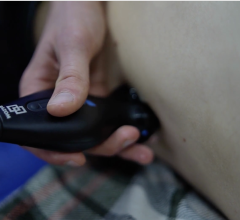
Ultrasound is increasingly being used outside the echo lab for point-of-care cardiac assessments in the office setting. Most of these systems are smaller, less expensive ultrasound systems used to triage a patient to determine the need for more detailed imaging exams.
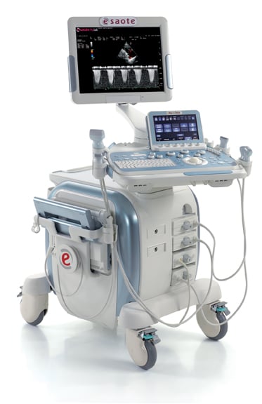
Smaller, lighter, portable ultrasound units are finding utility for remote telecardiology programs in rural areas. The echoes can be recorded at remote clinics and sent via secure websites to a central hospital to be read by an echocardiographer. Esaote promoted this concept in its booth at ACC.13, featuring presentations by cardiologists from Iowa Heart Hospital, which created a rural telecardiology program using Esaote’s portable systems.
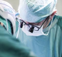
Transesophageal echo (TEE) is seeing rapidly expanding use in the cath lab and hybrid operating room (OR) to better visualize and navigate devices inside the body for increasingly more complex interventional procedures. The use of 3-D echo will likely see the most demand in procedures such as transcatheter ventricular septal defect (VSD) closures, left atrial appendage (LAA) closure and MitraClip mitral valve repairs.
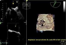
While 2-D echo remains the standard, there is increasing adoption of 3-D/4-D echo systems. These newer systems can allow better visualization, such as viewing the entire left ventricle, which users say is more accurate for quantification and measurements. The use of 3-D echo also offers surgical views that often aid procedural planning or interventional navigation.
Interventional Echo
A new trend in cardiac ultrasound is the rise of interventional ultrasound in both the hybrid OR and the cath lab. As minimally invasive and transcatheter procedures become more complex, there is a rising demand for more detailed, live anatomical imaging. Both transthoracic echo and TEE are seeing increased use for detailed procedural navigation (especially with 3-D and 4-D surgical view imaging) and to reduce radiation dose from use of angiographic fluoroscopy.
New Technology
To better optimize cardiac resynchronization therapy (CRT) lead placement, Toshiba has developed Activation Imaging as a new addition to its 3-D wall motion tracking software. The U.S. Food and Drug Administration (FDA)-cleared software is available on the Aplio Artida cardiovascular ultrasound system. When the heart is not functioning properly, different segments activate at different moments in time. Activation Imaging, in conjunction with 3-D wall motion tracking software, allows clinicians to evaluate dyssynchrony at the onset of the heart’s contraction and to properly identify the left ventricle’s pumping strength and timing for more accurate lead placements in CRT treatment.
Toshiba introduced new cardiovascular (CV) versions of its Aplio 500 and Aplio 300. Compact and easy-to-use, Aplio 500 CV and Aplio 300 CV offer exceptional image quality for 2-D cardiac exams and feature Toshiba’s 2-D wall motion tracking technology. The wall motion tracking software provides high-quality visualization and quantitative analysis of myocardial wall motion with accuracy and reproducibility. The system has on-board cardiac quantification measurements in all directions, including radial, circumferential, 2-D rotation and longitudinal.
Additional cardiac-specific technologies include tissue enhancement, advanced dynamic flow, lateral gain controls, tissue Doppler, stress echo, flex-M mode and auto IMT. Both systems are easy to use, with thoughtful ergonomics and a smaller footprint. The system offers three-click quantification measurements for strain, strain rates or ejection fraction.
Siemens demonstrated release 3.0 of the Acuson SC2000 volume imaging ultrasound system at ACC.13. It offers fully functional 2-D and volume imaging, and expands the usability of the system into pediatric echocardiography applications. It offers a neonatal transducer, with significant improvements in color Doppler and true fidelity color Doppler processing. The system also features a small transesophageal transducer optimized for pediatrics and neonatal imaging. Release 3.0 also expands Siemens’ portfolio of knowledge-based automated applications. The eSie Left Heart Measurement Package enables accurate, reproducible measurements of the left ventricle and left atrium. Siemens’ Native NTEQ ultrasound technology is a real-time technique that continuously optimizes image parameters that were previously unavailable for operator adjustment, enabling improved image quality without the need for additional steps, and encouraging efficient, intuitive and automated optimization of 2-D images. The capture preview function of the Acuson SC2000 system enables the user to review a sonography clip before storing it to an exam report.
Also showcased for the first time at ACC was the Acuson X700 ultrasound system. It is fully featured for TEE and intracardiac echocardiography (ICE) imaging, and comes standard with many advanced imaging technologies previously available only on Siemens’ premium Acuson S family of ultrasound systems, including micro-pinless (MP) transducer technology. Its 5 MP transducer system is designed to offer higher signal fidelity and improve signal-to-noise ratio, enhancing signal quality.
In addition, Esaote displayed its new eHD technology that refines each step in the imaging path. Transducer and beam former refinements extend depth penetration to aid in imaging larger patients. Other refinements increase contrast, reduce noise and enhance Doppler sensitivity to improve image readability. The company also displayed its XStrain 4-D technology, which offers a cost-effective method of improving the accuracy of quantitative data based on standard 2-D echo, instead of using more expensive volumetric transducers used for 3-D strain imaging.
The sonographer acquires standard, high temporal resolution 2-D views, and the system automatically creates a continuous motion model of the heart in 3-D space. Intuitive software allows cardiologists to interact with 4-D data and readily assess cardiac function, bringing economical myocardial quantification to the point-of-care.
SIDEBAR:
ASE Defines Focused Cardiac Ultrasound Practice Standards
The American Society of Echocardiography (ASE) released a new expert consensus statement focusing on the practice of using ultrasound as a tool for improving patient physical examinations. “Focused Cardiac Ultrasound: Recommendations from the American Society of Echocardiography” appeared in the June issue of the Journal of the American Society of Echocardiography (JASE).
The recent improvements in portability of cardiovascular ultrasound equipment have had a significant impact on its use in medical practice. As such, this new recommendation aims to inform the medical community and define how focused cardiac ultrasound (FCU) can be used to positively impact patient care. Technological advances have resulted in a growth in cardiac ultrasound images being procured by a variety of specialists and generalists in a range of settings, such as outpatient clinics, inpatient wards, critical care units, emergency departments and remote clinics. With the creation and publication of these recommendations, ASE is identifying how practitioners with limited training can appropriately use portable devices to expedite and improve the quality of patient care.
“The development of small, inexpensive, portable ultrasound devices, combined with a growing interest of physicians in many specialties to use ultrasound at the bedside for point-of-care assessment, prompted the American Society of Echocardiography to develop this expert consensus document,” said Patricia Pellikka, M.D., FASE, a cardiologist at Mayo Clinic and president of ASE. “This document establishes definitions for focused cardiac ultrasound, describes its appropriate application and discusses issues of training for the user. It will be a valuable reference for ASE members and for the medical community at large.”
This document will change practice for clinicians by encouraging additional training in light of the significant role the comprehensive transthoracic echocardiogram (TTE) has in the proper care and treatment of the heart patient. Key information obtained during the TTE is crucial in initiating proper therapies and in the evaluation of abnormalities. FCU is a focused examination of the cardiovascular system performed by a physician using ultrasound as an adjunct to the physical examination to recognize specific ultrasonic signs that represent a narrow list of potential diagnoses in specific clinical settings. The document distinguishes the emerging field of FCU as a bedside adjunct to the physical examination and echocardiography. Defining the distinctions between these techniques will allow practitioners to realize the utility of FCU while maintaining the value of echocardiography.
“Recent advances have resulted in the development of small platforms which have brought the power of ultrasound to the bedside,” said Kirk T. Spencer, M.D., FASE, cardiologist at the University of Chicago and ASE’s Guidelines and Standards Committee chairman, who served as the lead author of the guideline. “Physicians from diverse specialties, who have less training in cardiac image acquisition and interpretation compared to those trained in echocardiography, can learn to acquire and interpret cardiac ultrasound images as an adjunct to their physical examination assessment in clinical settings relevant to their scope of practice.”

 May 05, 2026