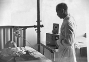
Dr. Tyra T. Hutchens (SNM founder) performs a radioactive Iodine thyroid uptake in 1953 using a Bismuth cathode Geiger Mueller tube. Photo courtesy of the Society of Nuclear Medicine.
As imaging modalities developed in the past 50 years — and ancillary products and services with them — colleagues who shared an interest in specific areas began to form groups to address the needs of their particular niches. Their focus included segments such as radiation oncology, nuclear imaging, women’s health, administration and informatics. The informal groups grew to become formal, well established organizations that contributed a great deal to the industry and today are a vital part of the imaging community.
Imaging Technology News asked a few of the imaging-related trade associations to share thoughts about what developments have made the greatest impact during the past five decades. They offered the following perspectives, as well as some background on their own histories.
AHRA: The Association for Medical Imaging Management, founded in 1973
According to our members, the most significant development in imaging technology in the last 50 years has been computed tomography (CT), followed by picture archiving and communications systems (PACS) and magnetic resonance imaging (MRI).
The imaging industry has dramatically shifted in complexity in this time, and the proliferation of technology has been led by a demand for increased efficiency and safety. These developments have taken place under the influence of economic pressures to limit healthcare costs and the increasing role of third parties (i.e., payers, regulators and government) in standard healthcare transactions.
Managing these evolutionary developments in the imaging industry has required continuous education and significant networking among industry professionals. AHRA, since its inception 40 years ago, has engaged this community and has provided a leadership and management vehicle for progressive learning, constructive leadership and synergistic networking to grapple with these dynamic changes. Members have been committed to and inspired by the organization’s meetings, publications and online opportunities for both education and networking.
SNM: The Society of Nuclear Medicine, founded in 1954
There has been much advancement in nuclear medicine and molecular imaging over the past 50 years. The expanded use of radiotracers labeled with Tc-99m and the development of single photon emission computed tomography (SPECT) and positron emission tomography (PET) have revolutionized the field. More recently, advances such as optical imaging, magnetic resonance spectroscopy and more have paved the way for new molecular imaging research.
The SNM was formed in 1954 to promote the science, technology and practical application of nuclear medicine. Since that time, the society has grown to 17,000 members — physicians, technologists and scientists specializing in the research and practice of nuclear medicine and molecular imaging.
Over the years, SNM has played an important role in creating forums within which these professionals can collaborate to improve patient care. The society has provided cutting-edge education through its meetings and has shared new research with its members through its influential journals. With these initiatives, SNM has improved healthcare by advancing molecular imaging and therapy.
ASTRO: American Society for Radiation Oncology, founded in 1958
“In the early 1960s, radiation oncology was just emerging as a clinical specialty. Fifty years later, radiation therapy is a well-established, essential component for curative and palliative treatment of malignancy,” said Michael Steinberg, M.D., FASTRO and ASTRO president-elect. “Recent advances in radiation therapy technology, such as intensity-modulated radiation therapy (IMRT), stereotactic radiosurgery (SRS) and stereotactic body radiation therapy (SBRT) allow for remarkable precision in treatment delivery with the realization of dose escalation and concomitant decrease in treatment-related morbidity.
“Image-guided radiation therapy (IGRT), critical to these modalities, incorporates advanced target imaging/localization utilizing ultrasound, stereoscopic shift imaging techniques, linear accelerator-based computed tomography (CT), and soon magnetic resonance imaging (MRI), to precisely guide radiation delivery,” Steinberg adds. “Image guidance for high-precision dose delivery, coupled with increasingly accurate tumor target delineation secondary to improved anatomical and biological imaging from advanced functional MRI and PET/CT, permit dose escalation, allowing increased tumor control probability and cures.”
ASTRO dates its origins to an informal meeting of the International Radiotherapy Club at Barney’s Market Club in Chicago on Dec. 15, 1955, said Gustavo S. Montana, M.D., FACR, FASTRO, chairman of the group’s history committee. This led to a formal meeting of about 60 people on Nov. 18, 1958, in Chicago, when the society was formally established as the American Club of Therapeutic Radiologists.
“Since then, the society has evolved, grown significantly, become international and become independent of parent organizations. To date, the society has more than 10,000 members,” Montana continues. “The specialty of radiation oncology as a primary or adjunct treatment modality for patients with malignant diseases has also evolved and is now a discipline based on scientific principles. The pretreatment evaluation, delivery of radiation therapy and followup of patients has become far more precise, more effective and safer since its inception at the beginning of last century.
“Being able to map out tri-dimensionally normal structures and tumor extent and evaluate treatment outcome is fundamental to obtaining the best radiation therapy outcome. This would have not been possible without the extraordinary advances in imaging technology,” he said.
AAPM: American Association of Physicists in Medicine, founded in 1958
Significant developments in imaging technology since the early 1970s have relied heavily on the phenomenal increase in computing capabilities, which led to the introduction of computed tomography (CT), digital radiography (DR), full-field digital mammography (FFDM), tomosynthesis, magnetic resonance imaging (MRI) and spectroscopy (MRS), positron emission tomography (PET), single photon emission computed tomography (SPECT) and advanced ultrasound systems, as well as computer-aided detection (CAD) and combined modalities, such as PET/CT, SPECT/CT and MR combinations.
Multi-detector, multi-slice imaging capabilities have sparked a transformation in imaging from the 2-D to the 3-D realm. For the X-ray technologies, major innovations in X-ray tube design (i.e., smaller focal spots, higher heat capacity and the introduction of new target and/or filter materials) have enabled higher resolution and contrast imaging with fewer artifacts. Greater computing power also lead to the combination of images and other data in radiology and hospital information systems (RIS, HIS), and greater image storage and distribution with picture archiving and communication systems (PACS).
AAPM’s formation in 1958 was “sparked by the confluence of two forces, one internal, the other external,” said one of those involved in the process, G.D. Adams, University of Oklahoma, Health Science Center, in an article he wrote for the association in 1978 (source: www.aapm.org). The external force, in a nutshell, was that an existing Hospital Physicists Association in England proposed forming an international organization. It was deemed desirable that such an organization, if formed, consist of societies and not individuals — and there was no such association in the United States.
The internal force, Adams said, “was the frustration experienced by physicists in biological and medical pursuits; in substance, we were not being recognized as competent professionals.”
“It seemed that physics in biology and medicine had no professional identity and that a national organization might speak for the profession in a louder voice than all the individuals, singly,” he added.
After a series of preliminary meetings, the association was formed in November 1958 in Chicago.
Another Group With Midwestern Roots — RSNA
The Radiological Society of North America (RSNA) is the behemoth organization covering all segments of the imaging industry. Founded in 1915, RSNA has more than 40,000 medical imaging professionals as members, including radiologists, radiation oncologists, medical physicists and allied scientists.
RSNA hosts the world’s largest annual radiology meeting, publishes two peer-reviewed journals, offers opportunities to earn CME, and provides research and education grants to young investigators.
It started as the Western Roentgen Society, but was renamed Radiological Society of North America Inc. in 1919. Through 1925, it held two meetings each year, with the winter event often held in Chicago. In 1926, it began having just one annual meeting, switching locations and frequently occurring near the Thanksgiving holiday or shortly thereafter.
In 1961, the event was held at the Palmer House hotel in Chicago. In 1975, it was held for the first time at McCormick Place in Chicago. It alternated venues in other cities a couple of times, but came to rest at McCormick for good in 1985.
The location of annual meetings that was a factor in the initial development of the group, according to RSNA’s history notes on its website. At the turn of the century, physicians interested in the potential of X-rays belonged to the American Roentgen Ray Society (ARRS), which was founded in 1900. RSNA says, “ARRS flourished throughout the first decade of the 20th century. But as the years passed, many radiologists who lived and practiced primarily in or near midwestern cities such as St. Louis, Chicago, Detroit and Cleveland began feeling ignored by the ARRS, because the ARRS continually held its annual meetings on the East Coast.
“The location of the ARRS meetings reflected the fact that the professional society had grown into an organization comprising mostly members from the Eastern states. Yet, in the early 20th century, this made the cost of travel to the ARRS annual meetings time-consuming and economically prohibitive for radiologists who lived west of the Appalachian mountains. In addition, many physicians believed ARRS membership requirements had become too restrictive.”
So in 1915, the Western Roentgen Society was born. More information about the development of the association and the industry can be found on the RSNA website, www.rsna.org.
SIDEBAR:
The Beatles and CT
Oddly enough, the annals of computed tomography history include a footnote about how the widely popular English rock group, The Beatles, may have had an unwitting influence.
Wikipedia notes that CT was originally known as the “EMI scan,” as it was developed at a research branch of EMI, a company best known today for its music and recording business. The first commercially viable CT scanner was invented by Sir Godfrey Hounsfield in Hayes, United Kingdom, at EMI Central Research Laboratories using X-rays. Hounsfield conceived his idea in 1967. The first EMI-Scanner was installed in Atkinson Morley Hospital in Wimbledon, England, and the first patient brain-scan was done on Oct. 1, 1971. It was publicly announced in 1972.
Wikipedia notes, “It has been claimed that thanks to the success of The Beatles, EMI could fund research and build early models for medical use.” The story is backed up by a 2007 online article by Whittington Hospital in London about a lecture given by Dr. Ben Timmis, consultant radiologist with the hospital.
It says, “As a direct result of The Beatles’ success, Dr. Timmis claimed, the scanner’s inventor, Sir Godfrey Hounsfield, was able to devote about four years developing the scanner from its 1968 prototype, to something that could be used in a clinical setting. His work was done in the Central Research Laboratory, a facility near Heathrow airport that was part of the EMI Group. Having sold 200 million of the Fab Four’s singles, (at seven inches, almost enough vinyl to stretch the length of the equator), the Beatles’ record company, EMI, was able to fund Hounsfield to do his research and the scanner was ready be used in hospitals in the 1970s.
“Dr. Timmis said that EMI’s research had initially estimated a worldwide need for only 25 of the machines, but thanks to their decision to invest in the pioneering technology, now there are thousands of the scanners worldwide being used in hospitals every day.”
Sources: www.wikipedia.com; www.whittington.nhs.uk
SIDEBAR:
MRI, a ‘Landmark’ in Imaging
Magnetic resonance imaging received a unique recognition earlier this year, when the American Chemical Society (ACS) designated the invention of MRI by Paul Lauterbur, Ph.D., as a National Historic Chemical Landmark. ACS is a nonprofit organization chartered by the U.S. Congress; it established the Landmarks program in 1992 to recognize seminal events in the history of chemistry and to increase awareness of the contributions of chemistry to well-being of society.
“Paul Lauterbur’s invention of MRI has transformed diagnostic medicine by providing a tool for the noninvasive, in vivo examination of soft tissues, such as the heart, brain and muscles, and for the early detection of cancer and other diseases,” ACS president Nancy B. Jackson, Ph.D., said. “It was the genius of Paul Lauterbur to realize that nuclear magnetic resonance signals could generate multi-dimensional images.”
The Landmark designation was celebrated at a March ceremony at Stony Brook University in Stony Brook, N.Y. Lauterbur began his work at Stony Brook University in the early 1970s by researching applications of nuclear magnetic resonance (NMR) imaging, an indispensible tool for chemical analysis. Lauterbur’s great achievement was to use the spatial information contained in NMR signals to make three-dimensional pictures. This led to a 1973 paper in Nature magazine announcing that he had generated multi-dimensional images of macroscopic objects. “A possible application of considerable interest…would be the in vivo study of malignant tumors,” he observed.
Lauterbur’s research was advanced by the British physicist Sir Peter Mansfield, Ph.D., Emeritus Professor of Physics at the University of Nottingham. The two shared the 2003 Nobel Prize in Physiology or Medicine for their discoveries concerning magnetic resonance imaging.
“Paul Lauterbur’s path-breaking work on magnetic resonance imaging has always been a source of pride for Stony Brook University,” said John Marburger III, vice president for research at Stony Brook University. “Now, the American Chemical Society’s declaration of this work as a National Historic Chemical Landmark raises the visibility of this achievement in a broader community. Most people know that MRI is a powerful medical diagnostic tool, but few stop to think how it came into existence.”

 June 24, 2026


