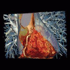
This coronary CTA clinical image shows exclusion of coronary heart disease. The image was acquired on a Siemens Somatom Definition - a dual-source CT system.
If “more is better” is the philosophy behind multislice CT (MSCT) technology today, then it isn’t surprising that 80 percent of the CTs installed in 2006 had 16-plus slices; and that trend will only multiply once Toshiba launches its 256-slice CT system in 2008.
So what is driving hospitals to buy up the slice chain? All signs point to the advanced cardiovascular applications performed on multislice CT scanners — in particular, cardiac CTA. Add to that the promise of an even broader range of applications in cardiac and brain imaging achieved with four times the slice capacity on a 256 CT system, and it seems like the more slices, the better.
But before hospitals go out and buy more slices, they need to take a closer look at what clinicians’ immediate needs are. Surely, surpassing 16 slices improves spatial and temporal resolution, but does expanding the volume coverage speed beyond 64-slices to 256 address clinical priorities such as better plaque characterization, greater visual detail inside stents and dose reduction? Some experts contend that wider coverage may overlook these acute clinical needs.
CTA: The Life Force Behind 64-Slices
Cardiologists are steadily joining radiologists in routinely using CT angiography (CTA) and MR angiography (MRA) for diagnosis. Over the next three years, CTA is expected to supercede SPECT and diagnostic catheterization procedures for certain cardiac conditions.
“It is the cardiovascular applications that are driving more slices. With the increased coverage of the detector, the inherent speed is providing real clinical benefit, specifically in imaging of the coronary arteries,” indicated Brian Duchinsky, global manager of CT product development for GE Healthcare.
“As a result, radiology departments and imaging centers are stepping up the pace of replacing their installed base and adding capacity. Going forward, the replacement market will range from 1,200 to 1,600 units per year over the next five years, and the total annual market will exceed 2,000 units,” said Lorna Young, senior director for Market Research, IMV Limited.
Many facilities are acquiring 64-slice CTs to expand their cardiovascular applications — in particular for cardiac CTA. This trend was further bolstered by a report presented at the 2006 American College of Cardiology that found that CTA rapidly and definitely excluded coronary artery disease as the cause of acute chest pain in less time, and at a lower cost, than stress imaging.
More Slices Means More Applications
Sixty-four slice CT is becoming the gold standard across radiology, cardiology and now in the emergency room. But just as clinicians begin to discover its advantages, clinicians are already looking toward the first 256-slice CT scanner, a system with capabilities that extend beyond cardiovascular applications.
Toshiba will soon introduce its 256-slice CT, currently a works-in-progress, designed to acquire a large volume of data that can cover the heart, brain and other organs in a single rotation with volume acquisition that reportedly provides more accurate images and lowers dose.
“The most significant advance that has been made possible by the 256-slice CT system is that it is now possible to perform whole-organ functional studies of the brain, heart, pancreas and other organs,” said Kazuhiro Katada, M.D., chairman of the Department of Radiology, Fujita Health University School of Medicine, Japan. “Such functional information can be obtained together with high-resolution anatomical information from the same dataset. Indeed, 256-slice CT is the first modality that can acquire functional data and anatomical data as a single dataset. Since these two types of data are obtained from the same dataset, they can be fused perfectly. It can be said that 256-slice CT provides full
integration of functional data and anatomical data.”
The 256 thus overcomes 64-slice CT’s limitation in capturing an entire image in a single rotation. In 64-slice CT systems, helical scanning must be employed, and datasets acquired over several cardiac cycles to cover the entire heart. Dr. Katada explained that when data for the entire heart is obtained by helical scanning, varying degrees of discontinuity in the body-axis direction are observed between the datasets acquired in different cardiac cycles. In addition, the acquired data corresponds to multiple phases after the injection of contrast medium.
“These problems are inherent to helical scanning, even when a dual-source CT system is used,” indicated Dr. Katada. “Research with the 256-slice CT, on the other hand, has provided data for the entire heart in a single rotation of the detector, acquiring data in the same phase of contrast enhancement and in the same cardiac cycle. This eliminates so-called “banding artifacts” in cardiac imaging.”
One of the most significant advantages in the acquisition of anatomical data is related to the improved accuracy of coordinate data. In 64-slice and earlier CT systems, helical scanning is employed and the patient couch must therefore be moved. In 256-slice CT, on the other hand, the need to perform helical scanning is eliminated, and it is not necessary to move the patient couch. This results in more accurate coordinate data, which is a great advantage when subtracting the data acquired before and after contrast enhancement. Dr. Katada also pointed out that “energy subtraction is another type of subtraction processing that is being researched. Previous research has indicated that satisfactory results cannot be obtained in energy subtraction if helical scanning is employed. Accurate energy subtraction is possible only in 256-slice CT,” noted Dr. Katada.
Another benefit of avoiding helical scanning is a reduction in exposure dose. “In helical scanning, scan overlap is unavoidable, but in 256-slice CT, when combined with prospective gating, such scan overlap is eliminated and only the required data is acquired. This leads to a significant reduction in the exposure dose,” said Dr. Katada. “Thus, 256-slice CT achieves remarkable improvements in anatomical data acquisition, but in my opinion, the most significant advantage of 256-slice CT is that it permits the accurate fusion of anatomical data and functional data.”
As neurologists know, “time is brain” in the case of stroke victims. A promising clinical application for CT256 is brain imaging of patients suspected of stroke. Dr. Katada describes how using his proposed scan sequence, in which 20 scans are performed intermittently, all of the required data can be acquired in a single session in 50 seconds. “A single session includes plain CT to detect the presence of early CT signs, subtraction CTA to acquire anatomical information concerning the vessels and perfusion CT to obtain tissue perfusion data. It is particularly noteworthy that when intermittent scanning is performed with X-ray conditions of 80 kV/80 mA, the exposure dose can be reduced to 5 mSv, which is dramatically lower than that in conventional perfusion CT studies. In addition, good low-contrast resolution is achieved by averaging the data in the time-axis direction. Research results indicate that the establishment of a quantitative cerebral blood flow evaluation method is expected in the near future,” said Dr. Katada.
Because 256-slice CT does not involve helical scanning, it reportedly permits accurate subtraction and the accuracy of coordinate data is improved. The most basic clinical application of subtraction is bone elimination. “The clinical advantages of being able to eliminate the complex bony structures at the base of the skull without difficulty and in a short time are immeasurable,” said Dr. Katada. “The data acquired in 256-slice CT studies, unlike the data acquired in MRI studies, contains bone data, and this bone data can be removed or added as required. This feature is extremely useful in the preoperative evaluation of lesions at the skull base and in the neck.”
“It is also possible to visualize the movement of the contrast bolus over time by performing subtraction between datasets acquired in different phases of contrast enhancement. Furthermore, 256-slice CT has great potential in other areas, including energy subtraction, as I mentioned earlier,” added Dr. Katada.
While Toshiba plans to commercially release the 256-slice system in 2008, it will install a beta system at The Johns Hopkins University School of Medicine and its Heart Institute in February 2007. Johns Hopkins will test the beta system for its value in early assessment of critical radiology and cardiac CT protocols. The beta system will be at the university for a limited time to acquire data to further clinical research and product development.
“The installation of the 256-slice beta system will be used by both radiology and cardiology and provides a tremendous opportunity to further medical imaging and research utilizing revolutionary CT technology,” said Business Unit Director Doug Ryan, Toshiba America Medical Systems.
Toshiba anticipates that the early adopters of the 256-slice CT will be major universities and research settings in large cardiovascular cardiology practices, as well as neuroradiologists performing complete whole-brain perfusion studies and for capturing the pancreas for diagnosing pancreatic cancer. The company also anticipates that, due to more than four million Americans having hepatitis C and needing liver transplants, radiologists will use the 256-slice CT for characterizing the liver.
Does 256 Meet
Immediate Needs?
A broad range of clinical applications delivered in a single CT system is a compelling argument for facilities considering an eventual upgrade to a 256-slice system. Some experts argue, however, that providing wider coverage by increasing the number of CT slices does not necessarily provide solutions to other more immediate clinical needs, such as improved characterization of plaque, greater visual details in stents and dose reduction.
“We don’t view more coverage with more slices as having a significant impact on where we need to go next — characterizing plaque and seeing details inside a stent,” said Duchinsky. “Simply by making the coverage wider, you haven’t enhanced those applications. What you will see from us are advancements in spatial resolution and the definition and a significant dose reduction.”
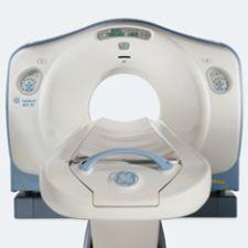
GE Healthcare believes it has responded to clinician requests with the introduction of the LightSpeed VCT XT, a system which reportedly captures images of the heart and coronary arteries in as few as five heartbeats and reduces patients’ radiation exposure by up to 70 percent for diagnostic cardiac scans.
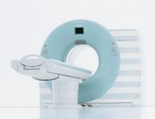
As an alternative to adding more slices, Siemens Medical Solutions introduced at the end of 2005 the Somatom Definition Dual Source CT (DSCT) scanner. The technology generates the high speed required for cardiac imaging by adding a second X-ray source and a second detector. The Definition is reportedly capable of scanning every heart regardless of heart rate without using beta blockers because of its heart-rate independent temporal resolution of 83 ms, and the system is said to deliver 50 percent less radiation in cardiac CT when compared to other single-source CT scanners on the market.
“It’s the fastest scanner in terms of temporal resolution on the market,” said David Roberts, M.D., of South Jersey Radiology Associates. “We had a Siemens Sensation 64, which is a standard single-source 64 detector system. The Dual Source Somatom is clearly better because of the two tubes that are mounted in the gantry. They nearly double speed with which you can capture images of any object, and what that means in the heart is that it’s able to create images of the heart so quickly that even if patients have very high heart rates, it can still create diagnostic images in that setting. So we get much, much less motion artifacts and a much higher image quality.”
The Definition recently received FDA clearance for several clinical applications, including: direct subtraction of bone in complicated anatomical regions; virtual un-enhanced liver images without multiple scans; evaluation of lung perfusion defects; visualization of cartilage, tendon and ligaments; and differentiation between hard plaques and contrast agents.
With the breadth of applications possible on Siemens’ DSCT, how significant are the advantages of the 256-slice system over a dual-source or 64-slice CT scanner?
What is clear is that “the 256 elements would allow for single-rotation imaging of the heart and reduce the image acquisition time,” Dr. Roberts said. “But what is unclear to me is whether the temporal resolution of that system will be superior to what we have now. Right now our temporal resolution is 83 milliseconds. If the new system offers a temporal resolution for single slice reconstruction that is below 83 milliseconds, then it is going to have a significant advantage. Otherwise, I don’t see the advantage because the challenges in cardiac imaging are really in getting rid of motion artifacts, and they aren’t necessarily in decreasing the breath-hold time. The breath-hold time for cardiac imaging is already at a very acceptable range.”
Managing CT Data Overload
An important consideration for buying more CT capacity is how to manage and archive increasingly large amounts of image data. A routine 256-slice brain or cardiac study has potential to generate up to 5,000 images, and up to 10,240 images for a more detailed neuro exam at a higher resolution, or up to 20,000 for a series of studies.
Prior to launching its 256 system, Toshiba is developing a data management solution in conjunction with partners Vital Images and McKesson to more efficiently handle such large data sets. “Before we bring the 256 to market, we have to be able to present a solution to clinicians for exactly that reason,” indicated Ryan. One approach Toshiba is working on with Integrating the Healthcare Enterprise is, “instead of storing slices, start storing volumes with a one header file in just a very large volume of data,” Ryan said, “making it more efficient to send over a PACS.”
The clinical advantages of the 256-slice CT seemingly outweigh any storage hassles — reinforcing the notion that the more CT slices the better. Dr. Katada’s vision for the future, however, dictates that what is best is the merging of multiple-source technology with CT.
“Ultimately, the ideal solution would be to combine all of these technologies. This may be possible within 10 to 15 years,” Dr. Katada noted. Right now what is essential, adds Katada, is to deliver a solution that provides the greatest benefit in all areas of the body.

 June 18, 2026


