Hardware and software advances are enabling echocardiography to greatly expand its capability with increased quantification accuracy, ease-of-use, increased workflow efficiencies and wider use outside of echo labs. Today, cardiovascular ultrasound systems are being integrated into point-of-care for triage, and in operating rooms and cath labs for procedural guidance to cut the use of contrast and ionizing radiation. Advances in 4-D echo are making it an enhanced tool for structural heart evaluation and visualization during procedures.
3-D, 4-D Echo Advances
3-D echo images a volume of data (similar to a computed tomography [CT] dataset) rather than the traditional 2-D image rendering. These volumes can be manipulated with advanced visualization software just like a CT, slicing images on any plane and enabling the creation of 3-D images that can be rotated.
The proliferation of 3-D echo was previously handicapped by the large amount of labor involved in creating images from a volume dataset, explained Stephen Little, M.D., FRCPC, FACC, FASE, cardiovascular imaging section, department of cardiology, Methodist DeBakey. He said earlier generation systems required 30 or 40 mouse clicks to create an image.
“3-D required a lot of manual processing to slice and dice the images. It just took too long to do anything,” Little explained.
However, he said the newer 3-D systems are making the technology more viable with automation. He said echo is following the same path previously followed by CT advanced visualization software, where automation made a big difference in its wider market adoption for daily use.
Two big technology innovations have recently made 3-D and 4-D systems more commercially viable for everyday use. First, there has been a rapid increase in computing power in less expensive, smaller packages. Second, the automation of many advanced visualization functions drastically simplifies use and reduces the staff time required to manipulate volumes.
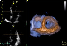
The introduction of 4-D echo (the fourth dimension is the addition of time) has opened new possibilities in ultrasound imaging. The analogy of 4-D is the difference between video and a still photograph. The technology allows 3-D images to be continuously updated for a live video view. The platforms with this feature require very fast processors to reconstruct large volumes of data into 3-D images over and over in milliseconds.
4-D ultrasound offers several advantages. It offers real-time color flow to assess hemodynamic information in the same heart cycle. It offers very accurate qualification of the left ventricle, free of geometric and shape assumptions used in 2-D echo. By using a 3-D volume of data, left ventricular wall motion tracking analysis can be done using the raw data volumes acquired. Vendors say this increases the accuracy of quantification.
It also offers multi-dimensional imaging, where operators can simultaneously acquire bi-plane and tri-plane images from the same heartbeat without moving the probe’s position. This offers two or three different axis views concurrently or as a composite view of the heart in real-time, offering a new field-of-view that previously could not be obtained. This helps acquire more information in fewer steps.
Real-time 4-D can produce images that are incredibly lifelike. This makes them easier to interpret and offers more meaningful information, including better procedural guidance. As technology continues to advance, 4-D echo will offer images comparable to CT 3-D reconstructions. Surgeons are now using 3-D echo reconstructions to aid procedural planning.
Use of 4-D greatly aids assessment of congenital heart diseases. Siemens recently introduced an updated version of its SC2000 cardiac ultrasound that quantifies volumetric color blood flow when evaluating holes in the heart (ASDs, VSDs, PFOs). The system uses a 3-D representation to show the true surface area and helps estimate the size of the holes for procedural planning.
Innovations in 4-D make possible real-time, comprehensive analysis of the beating heart during the entire cardiac cycle and allows even more detailed surgical-like views of the anatomy.
Toshiba’s new Aplio 500 shows the future of 4-D, where it can reconstruct volumes into color, fly-through video of vessel lumens. It works with peripheral vessels, but the heart is still too fast for the new technology to capture coronary vessels or ventricles. Image quality is similar to CT virtual colonoscopy.
Practical Application of 3-D
Methodist DeBakey Heart and Vascular Center has its own imaging center, which uses 3-D echo extensively. The center also images patients with both magnetic resonance imaging (MRI) and 3-D echo for comparative effectiveness research.
In the echo lab, 3-D echo is very good at estimating left ventricular ejection fractions (LVEF). However, there is a need for standardization between vendors before this technology will be used mainstream, Little said. Each 3-D echo machine is slightly different, so the workflow is not the same from vendor to vendor, and each requires use of proprietary workstations.
He explained 3-D offers a more accurate picture of cardiac function, but the basic concepts of 2-D echo still apply.
“3-D is not magic. It starts with a good 2-D image and you face all the same physics challenges as you do with 2-D technology,” Little said.
At DeBakey, echo contrast is often used to improve 2-D image quality when imaging obese patients, but they found 3-D has some limitations with contrast, said Miguel A. Quiñones, M.D., MACC, chairman, department of cardiology. The software uses automated 3-D tracking of the borders of the ventricle, he explained, but the automated tracking system is confused by the contrast and has issues. However, an operator can overcome this by switching to a manual mode.
Little said hospitals need to assess whether there is a need for 3-D. “It depends on what they plan to do with the system. If you plan to use it for surgical procedures, then it might be worth investing in a 3-D system. If you are involved in activities with more emphasis on structural heart, then 3-D has a lot of application.”
Expanding TEE Use
Little said DeBakey makes extensive use of 3-D echo transesophogeal echo (TEE) to better guide mitral valve prolapse and regurgitation repairs, atrial septal defects (ASDs) and trans-aortic valve repair (TAVR). In TAVR, he said TEE helps accurately place the angiographic pigtail catheter in the non-coronary cusp of the aortic root. It also offers Doppler flow imaging to evaluate the hemodynamics of the valves and check for paravalvular leaks.
Little explained 3-D TEE offers a definite imaging advantage during complex interventions. The use of an X-plane (also referred to as bi-plane) TEE probe allows visualization from two different angles. He said these views are displayed on the main screen in a cath lab or hybrid OR to better visualize where a catheter or device is located in the anatomy more clearly than 2-D angiography. This helps with procedural navigation and in cutting the radiation dose from fluoroscopy.
“You can get two views simultaneously from two different perspectives, which helps speed things up,” Little said. “It adds a level of confidence to show you where wires and devices are inside the heart.”
DeBakey uses 3-D echo from various vendors, including Philips, GE and Siemens, but only the Philips system had offered 3-D TEE, Little said.
Siemens recently introduced syngo FourSight 3-D TEE. It can scan the whole heart in one volume instead of stitching two or three images to create a whole-heart image.
GE Healthcare also has a new 4-D TEE system pending FDA review, which it previewed as a work-in-progress in March at American College of Cardiology (ACC) 2012 .
Comparison Chart
This article served as an introduction to the cardiovascular ultrasound systems comparison chart in the May-June 2012 issue of DAIC. To access the chart, click on the "comparison charts" tab at the top of the page. Participants included:
Esaote North America -www.esaoteusa.com
GE Healthcare - www.gehealthcare.com
Mindray - www.mindray.com
Philips - www.philips.com
Siemens - www.medical.siemens.com
Toshiba - www.medical.toshiba.com

 May 05, 2026