Regina Druz, M.D., FASNC, a member of the American Society of Nuclear Cardiology (ASNC) Board of Directors, chairwomen of the American College of Cardiology (ACC) Healthcare Innovation Section, and a cardiologist at Integrative Cardiology Center of Long Island, N.Y., explains the rapid expansion of telemedicine with the U.S. spread of novel coronavirus (COVID-19, SARS-CoV-2).
Druz spoke on the unprecedented expansion of telemedicine in the U.S. under COVID-19, seeing more use in the last two months, as opposed to the past two decades. The Centers for Medicare and Medicaid Services (CMS) previously only reimbursed for Telehealth in rural areas it determined had a shortage of doctors. However, in early March 2020, CMS dropped the geographic requirements and allowed Telehealth usage across th country as a way to mitigate person-to-person contact and keep vulnerable, older patients at home for routine check ups with doctors.
Druz has subspecialty certifications in nuclear cardiology, adult echocardiography and cardiac computed tomography (CT) and explains how Telehealth can be used to pre-screen patients and get patient sign off on procedures prior to coming in for an exam, helping speed the process in the hospital and limit personal contact.
Concerns about the rpaid spread of COVID-19 also has driven many radiology departments to convert to wider use of teleradiology to allow more radiologists to work from home and reduce person-to-person contact within the hospitals.
Watch the related VIDEO: Use of Teleradiology During the COVID-19 Pandemic — an interview with John Kim, M.D., chairman, Department of Radiology, THR Presbyterian Plano, Texas, and chief technology officer at Texas Radiology Associates.
Related COVID-19 Content:
VIDEO: Imaging COVID-19 With Point-of-Care Ultrasound (POCUS) — Interview with emergency physician Mike Stone, M.D.,
VIDEO: How China Leveraged Health IT to Combat COVID-19 — Interview with Jilan Liu, M.D., CEO for the HIMSS Greater China
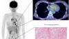
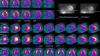
Study Looks at CT Findings of COVID-19 Through Recovery
Experts Stress Radiology Preparedness for COVID-19
ACR Recommendations for the Use of Chest Radiography and CT for Suspected COVID-19 Cases
VIDEO: What Cardiologists Need to Know about COVID-19 — Interview with Thomas Maddox, M.D.
The Cardiac Implications of Novel Coronavirus