McKesson's new Enterprise Image Repository, featured at the 2012 Society for Imaging Informatics in Medicine (SIIM) meeting, facilitates the sharing of images and data both within and outside an organization. It enables sharing of data with referring physicians and also among different departments, all at an affordable cost.
itnTV
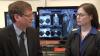
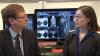
VIDEO: One on One with Amy K. Patel, MD, American Association for Women in Radiology Immediate Past President
Breast Imaging | April 15, 2024
Don't miss ITN's latest "One on One" video interview with AAWR Past President and American College of Radiology (ACR) RAN and RADPAC Chair, Amy K. Patel, MD, discussing advocacy initiatives and innovations in artificial intelligence (AI) for breast imaging.
Dr. Patel is a breast imaging trailblazer and radiology advocacy leader. In this video, learn how radiologists can support key initiatives, ways AI is improving patient care, and more.
Related content:
Technology Report: Artificial Intelligence in Radiology 2021
VIDEO: Integrating Artificial Intelligence Into Radiologists Workflow
Information Technology
Enterprise Imaging | July 17, 2012
Retention Management is a new feature of McKessonâ??s Enterprise Image Repository (EIR), offering the ability to delete data that no longer needs to be retained. It provides the flexibility to set different parameters according to different policies and requirements. System Dashboard is a new tool in the EIR which helps administrators manage and monitor how the system is performing. These features were showcased at the 2012 Society for Imaging Informatics in Medicine (SIIM) meeting.
Enterprise Imaging | July 02, 2012

Visage 7 enterprise imaging platform features a fast, thin-client, server-side processing technology that delivers enterprise diagnostic and clinical viewing solutions for healthcare institutions seeking to accelerate and enhance the delivery of radiology services. Building upon its enterprise-class technology platform, Visage Imaging has strengthened Visage 7 in the areas of performance, usability, integration, workflow, automation and advanced clinical capabilities.
Information Technology | July 02, 2012

ITN Editorial Director Helen Kuhl talks to Katherine Andriole, Ph.D., FSIIM, program chair for the Society for Imaging Informatics in Medicine (SIIM) 2012 meeting, about current trends in imaging informatics, including topics such as social media, quantitiative imaging and mobile technologies. Dr. Andriole also discusses some of the topics that will be of importance in the coming year, including the continuing challenge of integration and the evolving role of the radiologist.
Information Technology | June 28, 2012

Paul Nagy, Ph.D., CIIP, and Christopher Meenan, CIIP, discuss some of the opportunities available for Imaging Informatics Professionals (IIPs) at the Society of Imaging Informatics in Medicine (SIIM) annual meeting and through SIIM's programs and website — all of which are especially relevant in light of today's explosion of innovation in the imaging informatics arena.
Information Technology | June 27, 2012

J. Raymond Geis, M.D., SIIM Chair, and Mitchell M. Goldburgh, Dell Healthcare and Life Sciences, discuss the "Corporate Leadership Circle," a partnership allowing vendors to communicate with SIIM members in expanded ways about new technology and to create vendor/provider collaborations to resolve technology challenges.
Information Technology | June 27, 2012

Elizabeth Krupinski, Ph.D., FSIIM, and David Brown, BSCS, CNMT, CIIP, discuss the new SIIM Knowledge Center, a specialized website housing new educational material, including the updated "Need to Know" series, as well as discussion forums and top 10 lists.
Breast Imaging | March 30, 2012

Gary Levine, M.D., program chair/incoming president of the National Consortium of Breast Centers, gives an overview of current trends in technology, diagnosis and treatment of breast cancer, and regulatory activity that will impact women's health.
Women's Health | March 30, 2012

Gary Levine, M.D., program chair/incoming president of the National Consortium of Breast Centers, discusses legislation regarding breast density at the 22nd annual National Interdisciplinary Breast Center Conference (NCoBC), held in Las Vegas in March.
Women's Health | March 29, 2012

Gary Levine, M.D., program chair/incoming president of the National Consortium of Breast Centers, discusses how breast centers can use social media to educate the public regarding breast health and their services at the 2012 NCoBC meeting, held in Las Vegas in March.
Women's Health | March 29, 2012

Gary Levine, M.D., program chair/incoming president of the National Consortium of Breast Centers, discusses the role of politics on women's health in an election year, during the 2012 National Interdisciplinary Breast Center Conference (NCoBC), held in Las Vegas in March.
Advanced Visualization | February 13, 2012

The Chicago Zoological Society's (CZS) Brookfield Zoo is the first North American zoo to use 3-D advanced visualization imaging technology. This video shows a video fly-through of reconstructed 3-D computed tomography (CT) images of an aardvark, Humboldt penguin and African crested porcupine. The zoo is using Web-based software from Vizua to create animal CT scan advanced visualization reconstructions. Read the related article.
SIIM | May 20, 2011

Eliot Siegel, M.D., FSIIM, FACR, who is on the Board of Directors of the Society for Imaging Informatics in Medicine (SIIM), gives an overview of several programs that are part of the 2011 SIIM annual meeting and exoplains the benefits of the society. Siegel is professor and vice chair radiology for the University of Maryland School of Medicine Department of Diagnostic Radiology and chief of imaging for the VA Maryland Healthcare System.
Among the programs Siegel describes are the opening session on meaningful use rules; the "Ghost of Radiology Future," a look at the future challenge of regulations on the industry, and "Mind the Gap," about managing healthcare reform with informatics. The meeting also includes several new learning tracks, including ones on image sharing, improving efficiency in the reading room and imaging department, and new archival and storage models. There also will be round table sessions and a Sunday morning hands-on "Tools of the Trade" series.
For more information: www.siim.org
SIIM | May 20, 2011

Eliot Siegel, M.D., FSIIM, FACR, who is on the Board of Directors of the Society for Imaging Informatics in Medicine (SIIM), discusses a number of the society's initiatives and benefits of membership with ITN Editorial Director Helen Kuhl. Siegel is professor and vice chair radiology for the University of Maryland School of Medicine Department of Diagnostic Radiology and chief of imaging for the VA Maryland Healthcare System.
Siegel elaborates on a number of benefits of SIIM membership, which extend beyond the society's annual meeting. These include: the TRIP initiative, a workflow efficiency aid; the Imaging Informatics Marketplace buyer's guide; a mentoring program, and access to numerous resources on the website. SIIM also is considering several exciting initiatives for the future, including webinars and regional meetings.
For more information: www.siim.org
Radiology Business | March 22, 2011

Paul Chang, M.D., professor of radiology, vice chair of radiology informatics and medical director for enterprise imaging, University of Chicago, is a lead investigator on a closed-loop imaging research study that looks at all stages of imaging to optimize the imaging system at a hospital. The goal of the Philips-sponsored trial is to reduce errors and improving quality care and outcomes. He said it is important to optimize all stages of the imaging process. Chang explains the process they used for reviewing efficiencies and inefficiencies in the radiology department.
Watch another interview with Chang in the 2019 VIDEO: How Hospitals Should Prepare for Artificial Intelligence Implementation.
AAPM | March 22, 2011

American Association of Physicists in Medicine (AAPM) President Mike Herman, Ph.D., radiation oncology medical physicist, Mayo Clinic, explains the role of the society and its goal to improve patient care. Activities include sharing the latest scientific research, developing best practices, education, setting guidelines for certification and the roles of various staff under mediacl physicists, and how physicists can better serve their hospitals. The main focus in sessions at the AAPM annual meeting include patient safety concerning radiation dose and how to lower these doses in practice. Herman said AAPM is also calling for a national patient safety event recording process to make it easier to see where there are mistakes so they can be addressed. The society is also Herman said the process needs to be easy to access and use. He spoke to ITN at the some 2010, 52nd annual AAPM meeting
SIIM | March 22, 2011

Imaging Technology News Editor Cristen Bolan talks with Society for Imaging Informatics in Medicine (SIIM) Chair Bradley Erickson, M.D., Ph.D., Mayo Clinic, about how this year's annual meeting provides a glimpse into the future of imaging informatics. At the SIIM 2010 annual meeting, SIIM rolled out new interactive programs, such as "Doctor Is In" and the "Vendor Tie-In" sessions, to address problems with the application of information technology in the clinical setting.
The show's Opening General Session set the stage for the first annual Year in Review. SIIM's board of directors and other invited members were asked to identify hot topics for this session. The review covered the following topics:
• Politics and business intelligence: Meeting the challenge, as more administrators and radiologists are asked to justify expenditures and new equipment acquisitions while growing their imaging businesses.
• Advanced image visualization, quantitation and morphometric measurement of normal and abnormal structures: These tools are important, especially when using web clients and engaging clinicians has emerged as a high interest topic.
• Communication of patient data, 3-D, computed tomography and magnetic resonance processing, digital radiography, cloud-based storage: These examine aspects of healthcare reform and the role of informatics.
• Communication tools: Advancements in tools for communicating critical results.
Erickson said that SIIM is committed to providing more of this type of content radiologists are looking via the web. As Erickson looked toward the exciting developments in imaging informatics in the coming year, he had a take-home message for all of the attendees at SIIM 2010. "SIIM is the home for informatics professionals to learn what is new and exciting in the field and to establish connections with new people and refresh the connections they have established in the past," he said. "Having that sort of a network is critical. The annual meeting is a platform to establish connections amongst SIIM members throughout the year."
For more information: www.siim.org
Teleradiology | March 22, 2011

"Most people have no idea what a tremendous impact radiology and telemedicine have on poor and remote regions of the world," said Rebecca Cornelius, M.D., professor of radiology, neuroradiology, department of radiology, University Hospital, University of Cincinnati, College of Medicine. Cornelius was one of the physicians on the panel and video presentation "Zero Footprint Radiology and Telemedicine Build a Platform for Sustainable Care," which Imaging Technology News (ITN) hosted at the SIIM 2010 annual meeting.
The panelists described how physicians based in the United States used teleradiology and telemedicine technology to treat patients located in a remote clinic in Honduras. The panelists made the case that this technology suite is the basis for sustainable health care outreach programs in the future. ITN Editor Cristen Bolan then presented a video illustrating how physicians and technicians equipped The Roy and Melanie Sanders Frontera Medical Center in Honduras with the digital imaging and informatics infrastructure.
Several providers donated the suite of imaging technology. The equipment included a telemedicine system and ultrasound probe from Global Media, the VirtualPACS Web-based picture archiving and communication system (PACS) from MedWeb, a portable digital x-ray system from MinXray and a computed radiography (CR) unit from iCRco.
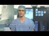
In this video, Dr. Juan Vasquez gives a live demonstration of how the imaging suite quickly and seamlessly operates. Vasquez started by taking an X-ray image, processing and reviewing it on the CR, and uploading the data set to the PACS in under 10 minutes. The guest of honor, Honduran Minister of Health Arturo Bendaña, himself a trained physician, easily toggled through the streamlined digital workflow. Vasquez explained how the transition from film to digital x-ray would save the clinic on significant costs incurred from developing film. Vasquez then examined a patient's thyroid gland with the ultrasound probe connected to a laptop computer. Next, he used a high-definition telemedicine camera to capture superficial anatomical images. Finally, he uploaded the images and consulted with physicians over Global Media's video-conferencing system. Jeffrey E. Heck, M.D., executive director and founder of Shoulder to Shoulder, explained to onlookers this was a model for delivering high-tech care, including expert specialty consultations, to some of the most remote and isolated areas of the developing world.
"With the addition of this technology, poor people have access to the same set of services that any well-equipped health center in the United States has access to," Heck said.
The panelists included: - Rebecca Cornelius, M.D., professor of radiology, neuroradiology (Clin Geo), University Hospital; University of Cincinnati, College of Medicine; Department of Radiology - Phillip Silberberg, M.D., head of Shoulder-to-Shoulder Radiology, pediatric radiologist, Kosair Childrenâ??s Hospital, - Roland Talanow, M.D., Ph.D., department of radiology, The Cleveland Clinic - Hayley Holland, MPH, director of grants and projects, Shoulder-to-Shoulder - Kim Guevara, corporate philanthropy officer and director of emergency management, Medweb. For more information: www.shouldertoshoulder.org
Related Radiology and Telemedicine in Honduras:
Radiologists Without Borders: The Heart of Radiology
ACS, Teleradiology Deliver Modern Healthcare to Honduras
Digital Imaging Delivers Modern Medicine to the Developing World
Digital Imaging Transforms a Nation
Radiology IT Explores New Frontiers
Computed Radiography Delivers Modern Medicine to Remote Regions
Radiology Delivers Modern Medicine to Rural Honduras
Angiography | March 22, 2011
Dr. Frederic Deschamps of the Institut Gustavy Roussy, France, explains his use of the Innova TrackVision application to plan and guide needle trajectories during vertebroplasty and oncology procedures in the interventional lab under angiographic fluoroscopy.
Performing needle procedures in the interventional suite frees up your CT system and provides better access to the patient. However, under fluoroscopic guidance, it may be challenging and time consuming to find the right entry point and advance the needle to avoid critical structures.
TrackVision 2 provides live 3-D needle guidance during your procedures. It lets you advance the needle down a planned trajectory overlaid on live fluoroscopy, visualizing any deviations from the desired path.
Highlights of the system include:
• Support multiple trajectories.
• 3D trajectories are registered in real time to C-arm and table movements, field of view and Source-to-Image Distance in real time.
• Visualize patient motion with the bone anatomy overlay and correct it at table side.
• Send bull eye's view angle to the gantry in a single click.
Pediatric Imaging | March 22, 2011

There is no doubt that medical imaging procedures save lives. However, one size does not fit all. Because children are three to five times more sensitive to radiation than adults, and cumulative radiation exposure can have adverse effects, it is critical for doctors to lower radiation levels when imaging a child. That is why in 2007, the Society for Pediatric Radiology (SPR) initiated the Alliance for Radiation Safety in Pediatric Imaging. Not long after, the American College of Radiology (ACR), the American Society of Radiologic Technologists (ASRT), and the American Association of Physicists in Medicine (AAPM) joined the Alliance.
The Image Gently campaign is the Alliance's initiative to raise awareness for lowering radiation dose used in pediatric imaging. The aliance is actively working with imaging manufacturers to standardize dose assessment and display for children. Although disagreements about the accuracy of the risk models or the degree to which the risks of radiation are emphasized are ongoing within the medical community, the message of the Image Gently campaign is clear: Reduce or "child-size" the amount of radiation used when obtaining a CT scan in children. To child-size the amount of radiation used, Image Gently encourages doctors to ask their medical physicist to determine the baseline radiation dose for an adult for that site's equipment and compare that dose with the ACR Standards.
While these guidelines are clear, it is not certain how widely doctors have implemented these radiation-reducing measures to date. To gage the impact Image Gently on medical imaging practices, Imaging Technology News (ITN) spoke with Marilyn Goske, M.D., chair of the Alliance, and Neil Johnson, M.D., president of the Society for Pediatric Imaging, both practice at Cincinnati Children's Hospital.
ITN: How serious a risk does radiation imaging pose to children?
Dr. Goske: One of the first things we need to remember is when children have imaging it is being done for an indicated medical condition and for a benefit for that patient. That is really what the Image Gently campaign revolves around. Once a study if medically indicated it behooves all of us in pediatric imaging to promote radiation protection and try to lower the dose and still maintain the quality of the exam so that we get the diagnostic information that we need. We know from studies, particularly from the atomic bomb survivors in Japan, that if children receive radiation from a bomb blast such as that one, they are more sensitive to radiation. Now medical imaging is different as it's a different form of energy and quite diffrent in how it's given for the imaging test, but it's the best we have. The data from that tells us that we need to be overly cautious and conservative, and that if we are going to use this technology, we want to use it in the safest way possible.
ITN: How exactly is the Alliance standardizing dose assessment and display for children?
Dr. Goske: We are working together under the direction of Keith Straus, who is the medical physicist at Boston Children's Hospital, Mr. Tom Toth, who is the former chief physicist at GE Healthcare, and Stephen Vastaghat the Medical Imaging Technology Alliance (MITA). The four major CT vendors have signed on to come up with more standardized dose displays so that when we complete a CT scan and we look at the images on task and that we have the information we need to interpret the information more accurately. Under the current system the CT dose that is displayed, which is the CT dye volume and the DLC are based on 32-centimeter adult-size phantoms. So if the patient is on the table and is exactly the same size as the phantom, the dose display is reasonably accurate. But in our patient population where you have an infant who weighs 5 lbs., for example, the younger they are, the smaller they are compared to the size of the phantom, and the more discrepant the dose display is. According Mr. Strauss in a paper that he published, the dose display can be off by a factor of three. So we are actually underestimating radiation dose for those small patients. We are working with numbers to get those displays more accurate so that radiologists, radiologic technologists and medical physicists have a better idea of what our smaller patients are really getting in terms of radiation dose during CT scans and other imaging procedures. Dr. Johnson: It's a very simplistic but important idea that we give our patients the right dose. We use the analogy of flying. We all fly in a commercial aircrafts, so we take risks. But there is a huge benefit when we minimize the risk. What we are trying to do is minimize the dose of radiation to children. We are not trying to stop these scans when they are needed medically. We are trying to do them with the minimum dose possible.
Information Technology | March 22, 2011
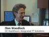
The HITECH Act, part of the American Recovery and Reinvestment Act (ARRA), and its impact on radiology is foremost on the minds of everyone in healthcare. Critical questions surrounding the language of the act remain unanswered. To gain better insight on the matter, Imaging Technology News spoke with healthcare IT research and development expert Don Woodlock, vice president and global GM of GE Healthcare Integrated IT Solutions.
Imaging Technology News (ITN): Will PACS and RIS qualify for reimbursement under the ARRA?
Don Woodlock (DW): The centerpiece of and the spirit of the HITECH Act is about adoption of general purpose EMR that go across the hospital or physician office EMRs for multi-specialty groups. The definition of meaningful use does mention images; all of the patient's test results have to be in the EMR, including images and imaging reports. Images need to be part of the electronic health record; [there is] mention of RIS/PACS, but it's not clearly spelled out that the stimulus will pay for RIS/PACS. I think the area where we need most clarity is in the outpatient- imaging environment. They are physicians, they see patients, and RIS and PACS is all they have, and they don't have another electronic medical record in that environment. Itâ??s my feeling that stimulus funds should be provided for physicians [who] use technology even though it isn't a traditional EMR.
ITN: On June 16th, the definition of Meaningful use? was released and included reimbursement for imaging described as multimedia (e.g. X-rays). A public comment period followed to assist Congress in clarifying this definition. What is the industry doing to represent radiology and convince Congress to include radiology's needs under the stimulus package?
DW: We are part of several groups that will provide feedback on helping Congress clarify this definition. We are part of Access to Medical Imaging Coalition, which is a group of imaging vendors. I talked to the chair of SIIM, Dr. Erickson from the Mayo Clinic and SIIM was going to get involved in defining meaningful use.
ITN: How will growing volumes of patient data impact radiology?
DW: There will be a big indirect effect on radiology. Radiology has been well automated for many years with RIS and PACS installed over the last decade. But they are basically working with physicians that have not automated at all, and I think the main impact that this Act will have is that there will be EMRs everywhere — hospitals and referring physicians will have EMRs as well. The way radiology interoperates and the workflow of the community will be a lot better when everybody has an EMR. A couple of examples are the radiologists [who] need the complete patient record to do a good job reading the patient exam. That includes patient history, problems, information about the order; patient allergies will be accessible to the radiologists in the click of the button. The orders will come in, in a cleaner fashion; right now they come in on paper, and radiology can help provide decision support in the ordering process, so that the right test is ordered for the right patient and the report will come in with all of the information that the radiologist needs. Then, inside radiology they will still use RIS and PACS to read and report on the exam, but then on the way back, the images and the reports will be embedded in the EMR so they will be widely available to every ordering physician that should have access. So the work of the radiologist will be more widely available to physicians that need it and the communication between the radiologists and the rest of the care team will be more effective once everyone is well-automated with IT systems.
ITN: Will the referring physicians be viewing all of the images on the EMR?
DW: That's right. [For] All the physicians outside of the radiology, their view of the world will be through the EMR. They will go through the EMR to see the full patient record including the imaging tests, the reports and will probably launch a browser to the images. So, we don't see the EMR becoming a PACS; the images will still be in the PACS, but there will be links to those images and Web browsers embedded in the EMR, so it will be easy for physicians to have access to this information.
ITN: Will the viewer in the EMR also have diagnostic capabilities?
DW: Probably not. In terms of a 6 mega pixel workstation, that will still exist in radiology. But these other physicians will have Web based tools and they may have access to diagnostic workstations and Web viewers, but that's not really what they are after. They want to see the images, and sometimes use 3-D tools, but they are not using it for primary diagnosis.
ITN: How will interfacing radiology PACS and EMRs impact workflow?
DW: The workflow will be much more streamlined. So, on the inbound side with the orders and the EMR information, we can eliminate paper with electronic order, we can make sure the right test is ordered.
© Copyright Wainscot Media. All Rights Reserved.
Subscribe Now