Technology | December 30, 2013

Tool helps take subjectivity out of breast density assessment

mammography systems women's healthcare vucomp m-vu breast density

December 30, 2013 — VuComp Inc. introduced M-Vu Breast Density, a tool for radiologists that provides automated assessment of breast density from a digital mammogram.
 
Clinical studies have shown that extensive breast density can make breast cancer detection in a mammogram more difficult and may also be associated with a higher cancer risk. Because of these issues, radiologists classify breast density using a four-level density scale established by the American College of Radiology (ACR) as part of its Breast Imaging Reporting and Data System (BI-RADS) standard. Patients who fall into one of the higher density categories may be recommended for additional screening exams. Legislation has been passed in 13 states requiring physicians to notify patients if their breast density is in one of the two higher categories.
 
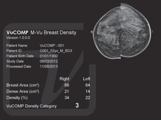
VuComp’s M-Vu Breast Density uses computer vision algorithms to evaluate the appearance of structures and textures in the breast to differentiate between fatty and dense regions. The algorithms then calculate a percentage of breast area that is dense and convert it to one of four density categories corresponding to the BI-RADS standard.
 
“In contrast to the volumetric approach, we designed M-Vu Breast Density to analyze the appearance of fibroglandular tissue rather than simply the total amount of such tissue, in order to better help doctors assess the risk that a cancer may be hidden in a mammogram,” said Jeff Wehnes, president and CEO, VuComp.
 
M-Vu Breast Density is immediately available. It can be combined with VuComp’s computer-aided detection system, M-Vu CAD, to provide a set of analysis tools for mammography.
 
For more information: www.vucomp.com

Related Content

News | Breast Imaging | Washington University

Jan. 22, 2026 — In breast cancer, a biopsy is the only diagnostic procedure that can determine if a suspicious lump or ...

Time January 29, 2026
arrow
News | Radiation Oncology

Jan. 27, 2026 — Researchers at the Icahn School of Medicine at Mount Sinai, in collaboration with other leading ...

Time January 29, 2026
arrow
News | Mammography

Jan. 16, 2026 — Vega Imaging Informatics has announced the successful curation of the world’s largest digital breast ...

Time January 19, 2026
arrow
News | Breast Imaging

Jan. 14, 2026 — VizMark has received U.S. Food and Drug Administration FDA 510k clearance for VM1, a non-metal visual ...

Time January 19, 2026
arrow
News | Breast Imaging

Jan. 14, 2026 — Susan G. Komen, the world’s leading breast cancer organization commends Assemblymember Lori Wilson (D ...

Time January 16, 2026
arrow
News | Breast Imaging

Jan. 14, 2026 — iSono Health has announced the commercial launch of ATUSA, an FDA-cleared, wearable and automated 3D ...

Time January 14, 2026
arrow
News | Women's Health

Dec. 12. 2025 — A new study has found that an individualized approach to breast cancer screening that assesses patients’ ...

Time December 17, 2025
arrow
News | Breast Imaging

Dec. 16, 2025 — Hologic, Inc, a medical technology company dedicated to improving women’s health, recently announced new ...

Time December 16, 2025
arrow
News | FDA

Dec. 8, 2025 — Lunit, a provider of AI for cancer diagnostics and precision oncology, has submitted a 510(k) premarket ...

Time December 08, 2025
arrow
News | Breast Imaging

Dec. 01, 2025 — DeepHealth, a wholly owned subsidiary of RadNet, Inc., has launched the DeepHealth Breast Suite,2 an end ...

Time December 04, 2025
arrow
Subscribe Now