May 30, 2013 — A recently published clinical study conducted at TOPS Comprehensive Breast Center in Houston, Texas, demonstrates that 3-D mammography (breast tomosynthesis) significantly reduces unnecessary recalls while simultaneously increasing cancer detection.
The study, "Implementation of Breast Tomosynthesis in a Routine Screening Practice: An Observational Study," was led by Stephen L. Rose, M.D., and published in the June issue of the American Journal of Roentgenology (AJR).
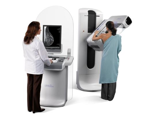
The study examined the use of Hologic's 3-D mammography in conjunction with 2-D mammography. The researchers found that 3-D mammography:
- Reduced recall rates by 37 percent;
- Increased the detection of cancers by over 30 percent; and
- Increased the detection of invasive cancers by more than 50 percent.
Instead of viewing all the complexities of the breast tissue in a flat image as with a traditional mammogram, 3-D mammography uses multiple low-dose images to create a 3-D view of the breast. 3-D mammography allows the doctor to examine 3-D breast images layer-by-layer.
The 2010 through 2012 study population included 13,856 women who received conventional 2-D mammography screening exams prior to the introduction of Hologic's 3-D mammography technology (breast tomosynthesis) and 9,499 women who received a 3-D mammography screening exam. Images in both time periods were interpreted by six radiologists with an average 12 years of reading experience.
"We are finding cancerous breast tumors as small as two to three millimeters," Rose said. "We are finding cancer earlier and this will allow us to reduce the amount of treatment patients need." Rose projects that as 3-D mammography becomes the standard breast cancer screening tool the size of tumors found will get smaller, and accuracy of deciding between benign and cancerous tumors will improve.
For more information: www.breasttomo.com
 October 09, 2026