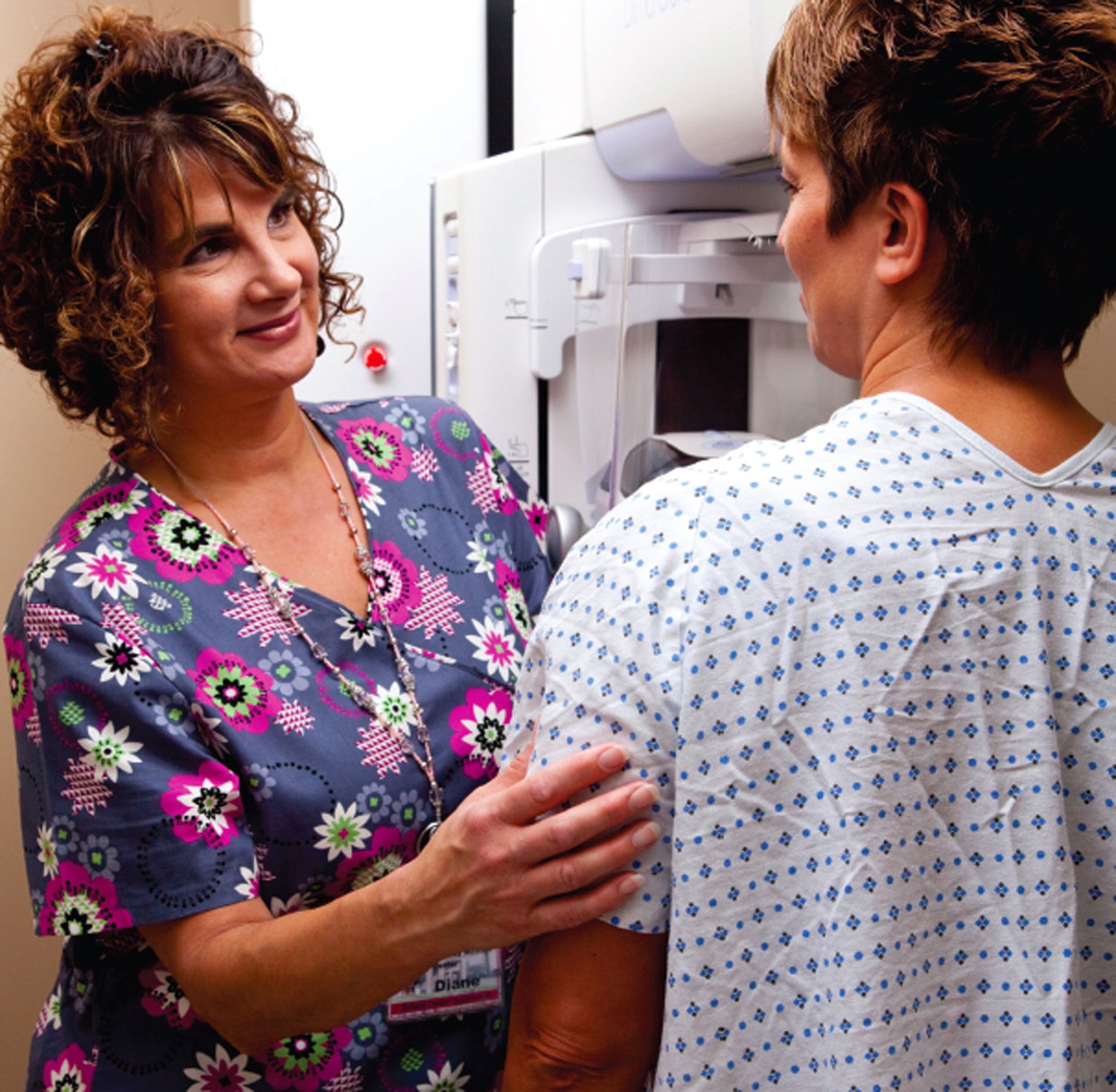
Diane Coady, R.T. (R)(M), breast center supervisor, talks to a patient about the 3D mammogram provided by Willamette Valley Medical Center.
“If I lived in a community without 3D mammography, I would strongly advocate my family go to a community offering the 3D exam,” states David Siepmann, M.D., radiologist at Willamette Valley Medical Center. A community hospital located 35 miles south of Portland, Ore., Willamette Valley Medical Center was the first breast center in the state to offer 3D mammography. In the first year, the hospital saw a 21 percent decrease in the overall recall rate and found a number of cancers it might have missed with conventional 2D mammography.
Willamette Valley offers 3D mammograms to all screening and diagnostic patients, finding the technology equally beneficial for both dense and fatty breasts. “We found 29 cancers on screening mammograms in the first year we offered 3D mammography,” states Diane Coady, RT(R)(M), breast center supervisor. “Eleven of those cancers were seen only on the 3D images. Eight additional cancers were seen best on the 3D images. It is mind-blowing when you look at these cases.”
Early in 2011, Willamette Valley was at full capacity with one digital mammography system and started looking for a second system to keep up with patient volumes. “We wanted a system that would not be outdated in a year or so, and we liked the ease of operation, resolution and clarity of the images on our Selenia system,” notes Coady. “So we looked at the Hologic Dimensions tomosynthesis system.”
Peace of Mind with Early Detection
“We’re saving patients the anxiety, cost and inconvenience of coming back for extra views,” observes Siepmann. “With 2D mammography, there was always some guesswork because of overlapping structures. With 3D mammography, you can separate out the layers of tissue. When you have a case that looks worrisome on the 2D mammogram, then look at the 3D images and immediately see it is nothing, it doesn’t take long to see the benefits of tomosynthesis.
“We’ve had a number of breast cancers that were not visible on the 2D mammogram but jump out as we scroll through the 3D images,” continues Siepmann.
Architectural distortions also appear more obvious on tomosynthesis images. “Many of the breast cancers found only with the tomosynthesis images were very small, less than 1 centimeter in diameter, which are the most important cancers to find,” notes Siepmann. “It is particularly exciting to find and treat small cancers before they can metastasize or cause trouble. Having the additional 3D information and that extra confidence in my interpretation makes a big difference for me.”
Patients are Excited About 3D mammography
As recalls decline with 3D mammography, Willamette Valley is filling the schedule with screening mammograms, experiencing a 10 percent growth in screening mammography compared to the same month last year. Some women are traveling from other parts of Oregon just for the 3D exam. “I’ve had patients travel four hours to have a 3D mammogram,” remarks Coady.
Siepmann concludes, “When you have a technology that improves the quality of care and reduces the overall cost of healthcare, there are very compelling reasons to adopt it. It is completely clear to those of us using tomosynthesis that it is a significant step forward. I am really pleased that we’re able to offer this amazing improvement to women in Oregon.”
Case study supplied by Hologic Inc.
The views and opinions expressed herein are those of David Siepmann, M.D., Diane Coady and their colleagues at Willamette Valley Medical Center and are not necessarily those of Hologic.
This information is intended for medical professionals in the United States and other markets and is not intended as a product solicitation or promotion where such activities are prohibited. Because Hologic materials are distributed through websites, eBroadcasts and tradeshows, it is not always possible to control where such materials appear. For specific information on what products are available for sale in a particular country, please contact your local Hologic representative or write to [email protected].
Hologic, Dimensions and Selenia, are trademarks and/or registered trademarks of Hologic and/or its subsidiaries in the U.S. and/or other countries.

 July 07, 2026


