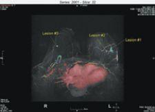
A malignant tumor, in three sections, involves the lateral aspect of the mid-left breast and the anterior aspect of the left breast.
The rapid advances in breast imaging techniques are making radiologists more accurate in diagnosing breast cancers and aiding surgeons in providing the best treatment options for patients. In fact, “seeing more” is forcing radiologists and surgeons to biopsy an increasing number of multiple lesions that ultimately lead to a more definitive diagnosis – and they are accomplishing the biopsies faster than the time it takes to perform stereotactic X-ray procedures.
“As we get more and more information from the imaging tools we have, we’re finding more disease,” said Randy Hicks, M.D., director of Breast Intervention and co-director of Breast MRI at Regional Medical Imaging in Flint, MI. “The long-term question is ‘Does that matter?’ and ‘Were the lesions always there and we just didn’t see them?’ We don’t know, but it’s truly amazing how many multiple lesions we find every day and that will intuitively lead to more biopsies.”
In most cases, Dr. Hicks said he’s performing multiple lesion breast biopsy using magnetic resonance imaging (MRI) faster than stereotactic procedures. Other studies are showing the same results.
“The beauty of MRI-guided breast biopsy is that you can image the patient and map out multiple biopsy sites,” Dr. Hicks said. “We load all of the biopsy coordinates at once, bring the patient out of the magnet and all I’m waiting for is the biopsy needle. The biopsy itself can be done in less than a minute. It’s the imaging sequences that take the most time.”
Regional Medical Imaging uses the MRI ATEC (Automated Tissue Excision and Collection) Breast Biopsy and Excision System (Suros Surgical Systems Inc.) with the grid method, a faster way to perform multiple lesion biopsies over the cumbersome pillar and post method, says Dr. Hicks. The time to biopsy a single lesion vs. multiple lesions is nominal, he said, with the only difference being the time it takes his technologist to swap a used biopsy needle for a new one to perform the second biopsy. Dr. Hicks averages a 22-minute total procedure time in MR breast biopsy.
In three MRI-guided breast biopsy studies using the Suros system, the median biopsy procedure time was between 33 and 38 minutes, including a successful biopsy completed in 17 minutes. Kara Carlson, M.D., medical director at Evergreen Breast Center in Kirkland, WA, said getting definitive information quickly and easily is good news for physicians and patients.
“Performing fast and accurate MRI-guided breast biopsies is so important for everyone involved, but most importantly for patients who need to know treatment options as quickly as possible,” she said. “After only a few procedures, we felt comfortable scheduling our MRI suite for a 60-minute time slot for multiple site biopsies.”
Evergreen Breast Center has seen its breast MR scans increase from six per month in 2003 to nearly 50 per month today. Biopsy procedures have seen similar growth, with the center performing an average of two per month in late 2004 to at least 10 per month now.
“Many of our breast MR findings were initially evaluated with an ultrasound exam,” Dr. Carlson said. With the difficulty due to positioning differences of the patient and in correlating MR findings to mammogram and ultrasound, we found ourselves going directly to an MR biopsy given the ease and simplicity of the exam.“
Dr. Carlson has biopsied four sites during one procedure, two in each breast, with a total procedure time of 70 minutes.
The industry is seeing an obvious shift in the acceptance of contrast-enhanced breast MRI and MRI-guided breast biopsy, particularly for those women with dense breasts or who are at high risk for breast disease or breast cancer staging. But even with the new acceptance of breast MR, average patient procedures still number less than two per month for those facilities equipped to do MRI-guided breast biopsies. Dr. Hicks and his associate David Strahle, M.D. perform 15-20 MRI-guided breast biopsies each month. Of their MR patients, more than 30 percent typically present with multiple lesions.
Other recent studies show even higher percentages of patients entering biopsy with multiple lesions. In a 2005 study by Constance Lehman, M.D., Ph.D. at the University of Washington Medical Center in Seattle, 50 percent of patients presented with multiple lesions, and in a 2003 study by Laura Liberman, M.D. at Memorial Sloan-Kettering Cancer Center in New York, 42 percent of patients revealed multiple lesion biopsy needs. At Evergreen Breast Center, 34 percent of patients revealed multiple lesion biopsy sites.
Drs. Carlson and Hicks say breast MRI will become more widely accepted. And while the current high costs associated with MRI may stall wide use of the imaging technique, Dr. Carlson said everyone will need to offer it to be competitive. More importantly, breast MR will allow earlier detection for mammographically occult breast cancers, she said.
“These are exciting times for radiologists,” said Dr. Hicks. “We are now able to answer that age-old question, ‘What is it?’ very quickly and very comfortably. Breast MR is giving us some shocking results. It changes the direction on what we are going to biopsy every day.”