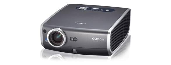
Canon's REALiS SX7 Mark II D Multimedia Projector.


March 29, 2010 - In the medical-education field viewing monochrome digital X-rays, computed tomography (CT) scans and magnetic resonance imaging (MRI) exams with accurate color and precise detail is of the highest importance.

A recently released projector is engineered to display medical imaging exams with precision in color and image quality. The monitors, the REALiS SX7 Mark II D, has a DICOM Simulation Mode that simulates the results of devices compliant with the Digital Imaging and Communications in Medicine (DICOM) Part 14 standardized display function for display of grayscale images. Because calibrating in a classroom, conference room or any other environment can be challenging when ambient light varies, this mode allows users to calibrate directly on the projector using 21 different levels of gamma adjustments. While not cleared or approved for medical diagnosis, this projector is designed for medical educators who need to display large images to properly train students and conduct lectures/conferences.
 
The SX7 Mark II D offers Canon’s LCOS technology combined with the projectors native SXGA+ high resolution (1,400 x 1,050) and 4,000 lumens of brightness. Small text can appear as small as 7 point.

Additional features on the project include:
 
- Canon 1.7x Powered Zoom/Focus Lens for ease of projector placement;
- DVI-I Terminal for digital connection for high-quality images and video;
- Quiet Operation produces only 31db (in Quiet Mode); and
- support of both Adobe RGB and sRGB color spaces.

For more information: www.usa.canon.com/projectors


Related Content

Feature | Artificial Intelligence

For the past decade, artificial intelligence's (AI) potential in healthcare has been synonymous with speed. In medical ...

Time February 16, 2026
arrow
News | ARRS

Feb. 11, 2026 —The American Roentgen Ray Society (ARRS) has announced the following radiologists, as well as their ...

Time February 13, 2026
arrow
News | Radiology Business

Feb. 3, 2026 — RadNet, Inc., a provider of high-quality, cost-effective outpatient diagnostic imaging services and ...

Time February 12, 2026
arrow
News | Digital Pathology

Feb. 11, 2026 — Leica Biosystems has announced the global launch of the Leica CM1950 Cryostat with DualEcoTec Cooling ...

Time February 11, 2026
arrow
News | Magnetic Resonance Imaging (MRI)

Feb. 6, 2026 — A state-of-the-art intraoperative MRI (iMRI) has arrived at the University of Chicago Medicine, one of ...

Time February 06, 2026
arrow
News | Computed Tomography (CT)

Feb. 4, 2026 — A new review published in the American Journal of Roentgenology (AJR) finds that advances in CT ...

Time February 04, 2026
arrow
News | Radiology Imaging

Feb. 4, 2026 — The Royal College of Radiologists (RCR) has issued its initial reaction to the British government's ...

Time February 04, 2026
arrow
News | Radiology Education

Jan. 22, 2026—The American Roentgen Ray Society (ARRS) will host a live virtual symposium, "Medical Imaging for ...

Time January 28, 2026
arrow
News | Computed Tomography (CT)

Jan. 21, 2026 — Aidoc recently announced that the U.S. Food and Drug Administration (FDA) cleared the industry's first ...

Time January 23, 2026
arrow
News | Point-of-Care Ultrasound (POCUS)

Jan. 22, 2026 — Qure.ai has received a grant from the Gates Foundation to develop a large open-source multi-modal ...

Time January 23, 2026
arrow
Subscribe Now