Carestream Health will debut upgrades to its newest PACS that include an embedded reporting module and a convenient graphic display for at a glance viewing of available patient records and data.
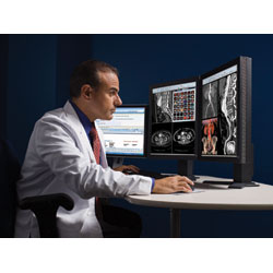
How do you define the ideal PACS workstation? There is no single answer just multiple solutions. Which one is right for your radiology department? That depends on the specific challenges facing your healthcare facility.
Nonetheless, there is a common thread of issues that create bottlenecks in even the most streamlined of PACS workflows. According to Gregory Pilat, RT, MBA, system director radiology/imaging services, Advocate Healthcare, Oak Brook, Ill., the key challenges are:
- increase efficiency in workflow to read more cases;
- disparate medical devices and IT systems;
- the trend toward declining reimbursement;
- the data tsunami;
- and hours of operation spread across several
time zones.
Reimbursement Woes
The existing complications in workflow are compounded by declining reimbursement and growth in data volumes, albeit incremental. In the post-Deficit Reduction Act (DRA) environment, radiologists are bracing for further cuts in imaging reimbursement from Medicare.
Despite a slowdown in the volume of advanced imaging since the implementation of the DRA at a rate of 1.5 percent 20081, Medicare spending on advanced imaging was reduced by 19.2 percent from 2006 to 2007 and volume of service grew by only 1.9 percent2.
Data collected by the Radiology Business Management Association (RBMA) and the American College of Radiology (ACR) highlights the imbalance between utilization and reimbursement. According to their recent survey, if reimbursements were reduced by half:
• 36 percent of practices would consider limiting access to Medicare beneficiaries
• 25 percent would consider dropping out of the Medicare program
• 40 percent would consider consolidatingservice sites
• 40 percent would consider closing their center
To compensate for lost reimbursement, the number of reads per radiologist must increase. But how will radiologists raise reading rates to handle a tsunami of data? First, PACS workflow efficiencies will have to significantly improve.
Big Bottlenecks
Some of the tightest bottlenecks in PACS workflow are caused by simple errors and IT functionality, which may involve: getting the image data to flow cleanly from the modalities to PACS; routing studies from PACS to the radiologist’s read station; making sure technologists provide accurate patient information when sending studies to PACS; monitoring all issues affecting the PACS directly, such as error messages, database errors.
When Frank Robbins the IT administrator for Next Generation Radiology, an outpatient imaging organization with four New York-based facilities, faced these very problems, he implemented the following solutions1:
- instantly routed all studies to radiologist’s read station to allow faster reads and increased radiologist productivity;
- routed images to the website, allowing referring physicians to examine images in their office in a short period of time;
- created interfaces to route studies from the PACS to the EMR both in house and to outside to referring physicians’ EMRs; and
- converted images to formats that could be stored on multiple EMRs.
Faster Reading Rates
One of the biggest problems with PACS is reading through large datasets.
“From a reading perspective, one of the largest challenges is the ability to diagnose large multi-slice studies that continue to grow in size and quantity while the pool of radiologists remains confined,” said Ron Muscosky, worldwide product line manager, Advanced Clinical Applications, Healthcare Information Solutions, Carestream Health. “Radiologists are finding the need to read more studies in a shorter period of time, so the difficulty lies with keeping up with this increasing amount of data.”
All too often, PACS providers develop solutions that add more steps. “Anytime you add a mouse click, scroll, page forward/backward, open/close, every step you add to workflow is a design defect,” stressed Pilat.
To speed up reading rates, radiologists need software solutions that automate and eliminate steps.
Features such as adaptive image-loading in McKesson’s Horizon PACS, can bring up large complex studies, while a three-click reporting feature eliminates delays in viewing images.
Other features that speed access to images are voice commands in RIS and, according to Muscosky, Carestream will soon provide the same and other navigational technologies in PACS.
Another strategy is to optimize the user interface to allow most commonly used tools to be most quickly accessible.
Next, there are shortcuts to reporting. “We see reporting becoming a much more seamless part of ones daily workflow. For example, the storing of structured reports can open up the ability for data mining that can be used for many purposes. Some of these include diagnosis based upon prior results and the availability of data for research purposes,” said Muscosky.
Some next generation features that eliminate mouse clicks and improve access to data include:
- touch screen
- voice commands in RIS and PACS
- automatic registration and matching of volumetric data at different time points on
- auto-structured reporting
- auto-loading templates mapped to procedure codes
- bookmark findings/optimal image
- one click to sign reports
- critical test results management solution to delivery of critical patient findings
- decision support and appropriateness criteria reference tools
Remote Access with i-Apps
Remote access also allows the radiologist to do his work at any time from any location. In addition to Web-based PACS, which is becoming a standard offering, there are other tools enabling remote access:
- iPhone App for 3D rendering of CT scans - app developed by Ziosoft (www.ziosoftinc.com)
- Osirix iPhone: The Osirix iPhone app is a DICOM viewer to the iPhone. (www.osirix-viewer.com/).
- ?eRoentgen Radiology Diagnosis: iPhone app with textual listing of recommended imaging studies from symptoms to diagnoses. (www.iatrossoftware.com)
- Microsoft Courier: Dual-screened tablet with multi-touch screen: designed for writing, flicking and drawing with a stylus, in addition to fingers.
Bridging Data Gaps
Other workflow issues involve patient information availability at multiple sites says Muscosky. “As the consolidation of facilities continue to occur, there is a need to effectively share data and balance the workload between them. In doing so, each site may have a different PACS solution already implemented. The challenge is sharing data between these disparate PACS systems at different facilities efficiently, so that a radiologist can read from anywhere with the same user interface.”
To bridge this data gap, Carestream has developed SuperPACS architecture to tie together disparate PACS systems allowing users to read from a single global worklist with the same user interface.
“The global worklist significantly aids in the balancing the workload by allowing exams at heavily loaded facilities to be read from less heavily loaded facilities without moving radiologists,” Muscosky explained.
Balancing Speed with Quality
With the inundation of cases radiologists are expected to read, maintaining quality can be tricky.
“Providing the radiologist with tools to optimize how they read exams or with tools that allow them to visualize exams in a more effective manner can play a vital role,” Muscosky pointed out. “For example, bringing 3D volume rendering into their normal reading can simplify and optimize the reading process, while simultaneously improving the diagnosis. The reason is that pathology, which may have been difficult to find or missed when viewing 2D images alone, may be much more obvious or visible on 3D volume rendered images due to the ability to observe and manipulate all spatial planes,” he said
A new viewing application, the PowerViewer by Carestream, provides automatic registration and matching of volumetric data at different time points and different modalities within a standard viewer. The PowerViewer allows a user to switch between different rendition types, such as MPR and MIP, including volumetric data in various planes. Automatic registration is designed to allow users to manipulate one dataset in any spatial plane and the other exams will follow.
By registering the images volumetrically, the user can quickly assess any change in pathology because the current and prior exams are fully synchronized in all spatial planes. For example, if a patient is being diagnosed for lung nodules, they may have a number of prior exams in addition to the most recent exam for comparison.
“Typically, one or more slices in the prior exam would have been marked as key images, or identified as having the most significant pathology. Additionally, the pathology may have been outlined with an annotation tool and saved,” said Muskosky.
“The radiologist would just need to quickly jump to the key image containing the outline pathology on the prior exam and the registered recent study would display the synchronized view. Our specialized Relate Tool allows users to locate the pathology on the prior and the application automatically identifies that location on the most recent and other datasets. Doing so allows the user to make a quality diagnosis very quickly with a minimal number of clicks,” said Muscosky.
Easy Access Decision Support
Another key to quality is easy access to evidence-based clinical decision support solutions. Web-based decision support tools, such as InterQual Online Anonymous Review by McKesson, enable quick reference to the clinical database, in this case InterQual Criteria, in a protected environment that doesn’t expose patient-identifying information.
Radiology-specific search engines also streamline access to decision support. A few examples include:
- Radiology Research
- MyPACS.net
- Yottalook.com and Yottalook app for the iPhone
- ACR Case in Point
- Dr. K. MSK Cases
- EURORAD
- Journal of Radiology Case Reports
- Pediatric Radiology
- Rad Files
- radRounds Cases
Getting the Message Across
Never underestimate the power of Murphy’s Law - if something can go wrong, it will go wrong.
After studies revealed hundreds of diagnostic imaging cases with critical results findings fell through the cracks,4 the American College of Radiology updated its guidelines with communicating critical diagnostic imaging findings,5 and the Joint Commission added critical results reporting guidelines to its National Patient Safety Goals.6
There are several approaches to delivering critical results.
“In many situations it is a PACS or RIS/PACS system that provides the communication of these critical results and critical results analysis. For example, critical results may consist of notes and key images added to the patient record in PACS. Reports can then be generated in PACS and/or RIS containing this information along with additional structured data through dictation and speech recognition that are then communicated to the responsible caregiver,” said Muscosky.
Robbins addresses this problem by routing images through the PACS directly to the doctors’ read station. This way, “We give our radiologists the ability to look at a patient’s exam literally before the patient is out of the scanning room,” said Robbins. “If a critical result is obtained action can be taken before the patient leaves the facility, if needed. It also decreases the time it takes to notify a referring physician of any critical results involving their patients.”
One option is through structured reporting. “The use of structured reports allows for consistent and accurate results,” indicated Muscosky. “Although the standards indicate critical results should contain critical or non-standard results, CARESTREAM RIS/PACS provides such results for all types of exams in a fast and thorough manner. The RIS or PACS reporting system can then use the data collected for analysis to determine if the expected results were met.”
A critical test result management (CTRM) tool, Veriphy from Nuance Communications provides a robust solution that includes: policy development and implementation; communications technology that verifies message receipt and automates documentation and rule compliance-user-friendly; live monitoring of report delivery; and real-time reporting that enables benchmarking and performance evaluation.
True Multimodality Means Mammography
Although breast imaging is an integral part of the radiology, progress in developing a truly unified multimodality reading environment that enables viewing all breast exams – mammography, ultrasound, MRI, and molecular images – has been delayed. More recently, vendors have been providing multi-modality workstations with integrated mammography and dedicated breast imaging capabilities.
Retrieving breast images is one of primary challenges for breast imaging at many hospitals. “There is a real need for a central data repository for all breast images and reports for mammography, ultrasound, MR, PEM, BSGI. We store by modality and not by the individual. We also need easy access to priors,” said Pilat.
At Clinical Breast Imaging at the University of Chicago, Gillian Newstead, M.D., director of the center, also underscores the problem with importing images from third-party vendors and having to readjust images for viewing. “It has not been an easy matter to import different types of mammography images onto different vendor workstations,” said Dr. Newstead. “The images may come over so that you can view them, but they may not line up properly or they are different sizes. There is a lot of manipulation that the radiologist has to do so that you can read them,” She added, “We also have a unique situation where we require special workstations for viewing mammography images; they have to have 5 megapixel (MP) viewers.”
Siemens Healthcare facilitates image imports with MammoReport, which enables the user to retrieve old films once they have been converted or “scanned” into a digital DICOM format. Another handy tool is the MammoDiagnost VU Workstation by Philips Healthcare. “The MammoDiagnost VU in fact does import quite well disparate types of mammography images and displays them in a similar fashion, which is an advantage,” said Dr. Newstead.
To facilitate image retrieval, Sectra’s ICS5/mx.net diagnostic radiology breast imaging workstation provides full PACS functionality, enabling the user to access the entire image chain - all priors and currents.
Dedicated mammography tools that should be available with an integrated breast imaging workstation should include:
- dedicated keypad for mammography workflow
- automatic image display and launch CAD for mammography
- user-defined viewing protocols
- advanced post-processing tools
- MPR built into the viewing protocols to display automatically without intervention of the radiologist
- web deployable architecture for upgrades
Enterprise PACS Integration
The integration of radiology PACS with IT systems across other specialties is inevitable and into the EMR, but it may require some adjustments to workflow.
“The most challenging aspect today is integrating the products seamlessly,” pointed out Muscosky. “Although standards based communication protocols exist, many systems may be homegrown and implement an interpretation of the interface.”
GE Healthcare addresses enterprise PACS integration with its Xeleris solution for nuclear medicine, which is integrated on the Centricity PACS workstation. Xeleris provides advanced imaging toolsets for the viewing and analysis of perfusion and metabolic pathways in SPECT/CT and PET/CT studies serving neurology, cardiology, and oncology departments. The Centricity PACS-IW Oncology Workflow also integrates sophisticated PET/CT fusion and SUV measurement capabilities into the web-based PACS-IW.
“With the existence of RIS, EMR systems, and other sources of data, another challenge being faced is how to provide the user with an effective means of presenting all available patient data, while not overburdening them so that their efficiency and interpretation is jeopardized.”
ScImage has come out with a toolset for delivering PACS content to electronic medical records systems (EMR) through the PicomWeb.Images, waveforms, final reports and other diagnostic documents are available to users of the EMR. Users can browse to patient images and data for clinical review and access using standard image manipulation tools, then switch to full diagnostic viewing with a single click.
“The result is the creation of a physician portal that delivers images, reports, waveforms and documents to EMR users, on demand,” said Sai P. Raya, Ph.D., ScImage Founder and CEO. “Importantly, the content and relevant tools can quickly transition to full diagnostic mode for those users with the right credentials.”
Wish List
The pressures of declining reimbursement will continue to hone the capabilities of PACS for improved workflow and increased productivity. But what might some of the future enhancements might be?
Muscosky points to radiologist productivity increases gains through features that enhance image presentation, thus provide access to studies on a timely basis through. He added, “By bringing up a study in any office in the practice allows better distribution of the work, again increasing productivity.
For Robbins, his wish list of future PACS features would include:
- automatic web integration of EMRs without use of VPNs.
- automated workflow of studies to EMRs;
- automated workflow on studies being sent out to outside hospitals for interpretation;
- notification via e-mail to IT personnel if anything within the PACS starts to fail.
To some these workflow enhancements may sound basic, but to a radiologist it can mean the difference between 100 and 200 cases a day.
References:
1. Trends in Imaging Services Billed to Part B Medicare Carriers and Paid under the Medicare Physician Fee Schedule, 1998-2008. THE MORAN COMPANY Executive Summary Prepared for the Access to Medical Imaging Coalition (AMIC) October 2009. 10/9/2009
2. Previous Moran and Company analysis.
3. Merge PACS.
4. Bates DW, Leape LL. Doing better with critical test results. Jt Comm J Qual Patient Saf. 2005 Feb;31(2):66-7, 61.
5. American College of Radiology ACR Practice Guideline for Communication of Diagnostic Imaging Findings. Practice Guidelines and Technical Standards 2005. Reston, VA: American College of Radiology; 2005. pp. 5-9.
6. Joint Commission. National Patient Safety Goals. Available at: http://www.jointcommission.org/PatientSafety/NationalPatientSafetyGoals…. Accessed January 28, 2009.
7. American College of Radiology ACR Practice Guideline for Communication of Diagnostic Imaging Findings. Practice Guidelines and Technical Standards 2005. Reston, VA: American College of Radiology; 2005. pp. 5-9.


 November 29, 2025


