September 29, 2009 - Philips Healthcare will present in its booth (2113) at the ASTRO 2009 annual meeting, held Nov. 1-5, at McCormick Place in Chicago, Ill., the following technologies as its comprehensive solution to image-guided radiation therapy (IGRT).
o 3D treatment planning is by definition image guided and Pinnacle was the first fully 3D treatment planning system.
o Image guidance starts with diagnosis and Philips offers the most comprehensive portfolio of imaging systems designed for Cancer Care – Brilliance BB CT, Panorama high field open MR system, the Gemini TF PET/CT and now the GEMINI TF Big Bore PET/CT featuring bore sizes of 85 cm in diameter.
o These same imaging devices are used for monitoring patient’s progress both during and post therapy.
o Respiratory correlated imaging on our imaging devices provides better image quality and supports all approaches for treating cancers of the thorax and abdomen where motion is a significant factor.
o Multi-modality image support via Pinnacle’s Syntegra automated image registration functionality in simulation and treatment planning ensures that the true extent of the disease is incorporated into the treatment plan, leading to better outcomes.
o Convenient 4D data handling and analysis tools in Pinnacle are available to seamlessly incorporate these data into the treatment planning process.
o Both the AcQSim3 Virtual Simulation Software and the Pinnacle3 Treatment Planning System can accept image information from all CT scanners and most diagnostic modalities, including on-board imaging systems on linear accelerators. Both systems accept gated and phased datasets from multi-slice CT scanners such as the Philips Brilliance CT and GEMINI Time of Flight PET/CT scanners for oncology. Being able to assess and manipulate this information allows physicians to gauge both inter-fraction and intra-fraction motion of tumors in their patients for extremely accurate target localization.
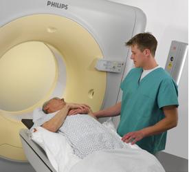
Both the CT and PET/CT scanners from Philips are available as large bore scanners, featuring an 85-cm bore size. This allows patients to be scanned in the same position they will be treated in, offering unrivaled imaging accuracy.
o Model Based Segmentation (MBS) provides automatic contouring tools to streamline critical structure definition and range of motion.
o Organ propagation via MBS provides near instantaneous complex re-contouring on image sets acquired during the course of therapy in order to assess the need to re-plan the patient (due to significant anatomical or physiological changes during therapy). If re-planning is necessary, it is much less burdensome given that the required contours are quickly generated based on the original planning CT dataset.
For more information, go to ITN's ASTRO FastPass site: itn.reillycomm.com/astro/