St. Peter’s Breast Center, located in Albany, N.Y, added 3D mammography (breast tomosynthesis) in 2011 because the staff knew it would improve diagnostic accuracy; even so, the technology has exceeded all expectations. In 2012 more than 22,000 mammograms were performed at the breast center.
Two-thirds of those studies, more than 14,000, were 3D exams.
Initially, St. Peter’s replaced one of its 2D units with a Hologic Selenia Dimensions 3D mammography system. Within a few months, they added a second unit to keep up with demand, and plan on transitioning their two additional systems to 3D in the near future. “We have a solid base of patients who benefit from the improved diagnostic capabilities of 3D mammography,” stated Andrew Warheit, M.D., medical director of the breast center and attending radiologist at St. Peter’s Hospital.
Since opening its doors a decade ago, St. Peter’s Breast Center has become the most preferred facility in the capital region for dealing with complex cases. The center has been named a Breast Imaging Center of Excellence by the American College of Radiology (ACR), and was the first center in the capital region to receive the national designation.
Improved Workflow, Better Diagnostics
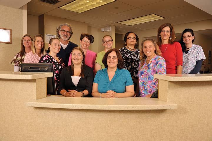
St. Peter’s Breast Center has six dedicated breast imagers and offers a comprehensive suite of breast health services including mammography, ultrasound, breast MRI and breast biopsies. The essence of the breast center is its combination of prompt care, expertise and the most modern, specialized breast-imaging technology.
“Our focus is to offer women a single location for all breast health services,” explained Elizabeth Malloy, breast center supervisor. “When a patient comes in for a diagnostic mammogram, we can perform an ultrasound and biopsy on the same day so they don’t have to keep coming back for additional testing. We can do this partly because 3D mammography is decreasing the number of callbacks and improving the efficiency of our workflow.”
The technology is making a significant improvement in the breast center’s diagnostic capabilities. This is particularly true for women with dense breast tissue, although Warheit notes all patients can benefit from a 3D exam. “For a fatty breast, 3D can rule out abnormalities,” Warheit said. “If you find little nodules or little round densities, you can look through the slices on the 3D images and see clearly the fatty hilum and you know it’s a lymph node right off the bat. In a dense breast, the underlying dense tissue melts away on a 3D image.”
Added Malloy, “It’s an amazing technology that is critical in diagnosing cancers at the very earliest stage.”
Fewer Callbacks Frees Up Resources for Diagnostic Exams
At first, the breast center predominately used 3D for diagnostic mammograms, only performing screening mammograms if the system had an open slot. But over time, more physicians and patients began requesting 3D.
Additionally, 3D created new efficiencies at the breast center, including fewer callbacks and more time for diagnostic exams. Warheit reports 3D mammography has cut the breast center’s callback rate by 50 percent. “With 2D, many patients were being called back
to evaluate asymmetries. We’re finding 99 percent of these asymmetries are easily clarified on the 3D images, identifying them as superimposed tissue or overlying densities rather than anything real.”
Preventing Unnecessary Biopsies
“I think the real benefit is not getting all the way to biopsy for something that turns out to be benign,” added technologist Darlene Pesnel. “Most biopsies do turn out to be benign; but, in the past, many patients had to get to tissue sampling for a definitive answer. With 2D, you would see little things that were vague, just innocent asymmetries, but no matter how you positioned, no matter what you did, you couldn’t make them go away. Now, 3D answers those questions right out of the gate. Plus, when we do find something that’s real and hidden in tissue we know exactly where it is, what depth it’s at, and what quadrant it’s in. You know exactly how to look for it with ultrasound or with MRI. So, we get the very best tissue sampling of the proper area and a fast diagnosis.”
More Physicians Are Specifying 3D Mammograms
The breast center sent a letter to all area physicians informing them of the availability of 3D and the benefits it offers patients. As a result, they began seeing referring physicians request 3D. “We are seeing providers write ‘3D Mammo’ on the script,” states Malloy. “That’s brand new. Now physicians understand what 3D can do for their patients; and, if they know someone has had a lot of difficulty, or is at high risk for breast cancer or has been called back year after year, then 3D is the right technology.”
“We don’t charge for the 3D exam or bill for anything additional on top of a routine screening or diagnostic mammogram,” concludes Warheit. “Early detection is our mantra, and the improvement in diagnostics is well worth the time. 3D gets better outcomes and it saves lives.”
Case study supplied by Hologic, Inc.