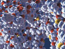
The molecular data visualized is from a large-scale simulation by the Theoretical Biophysics Group of the Beckman Institute at UIUC. These figures are images of a protein molecule, composed of approximately 20,000 atoms. These data come from a single timestamp of the simulation run on an SGI system. Images courtesy of the SGI institute.
As scientists focus on eradicating cancer, heart and neurological disorders, innovations in medical imaging and IT are bringing us closer to prevention, treatments and cures for these deadly diseases.
Considered by The New England Journal of Medicine “one of the most important medical developments of the past 1,000 years — ranking with the discovery of anesthesia and antibiotics,”1 medical imaging is the digital microscope that enables physicians to see inside the body and inside cells to screen, diagnose and stage disease; monitor treatments and disease recurrence; and facilitate medical research in areas such as drug discovery.
Many recent innovations in medical imaging might appear to be straight out of a sci-fi film. However, new technology that tracks a physicians’ subconscious eye movements as he or she reviews images could one day become the standard of care. The emergence of “smart” technologies integrating engineering and biological approaches has culminated into an influx of applications for FDA premarket approval for new imaging devices, many of which work with pharmaceuticals.
“The FDA is already seeing a steady increase in the number of requests from developers for presubmission meetings to seek advice on the best approaches for scientific and clinical testing and evaluation of cutting-edge technologies, such as molecular medicines (genetic and proteomic diagnostics and therapeutics) and products developed using nanotechnology.”2
Over the last 30 years, Imaging Technology News (ITN) has brought its readers the latest innovations and developments in medical imaging. As we look toward the future, ITN chose a select group of physicians, IT experts and its Advisory Board to rank, on a scale from 1-10 (1 having the most impact on medicine), what existing medical imaging and IT technologies will significantly enhance patient care in the next decade. The results were as follows.
1) Biomarkers for Drug and Imaging Development. Biomarker research in cancer diagnosis and drug development is years ahead of other indication areas. Biomarkers, which are indicators of a particular disease state or a particular state of an organism, used with imaging offers the potential of increased efficiency for drug development.
For example, researchers employ tumor uptake of 18F-FDG by PET scanning as an internal decision-making tool in its evaluation of drugs for the treatment of tumors, and examine the close correlation between this uptake and tumor regression using a CT or MRI scanner. The use of biomarkers in imaging is expected to validate and apply imaging-related biomarkers at all stages of drug and medical device development.
2) Targeted Imaging, Targeted imaging combines a contrast agent with an adhesion molecule to target the contrast directly to the desired region. Site-specific adhesion molecules such as monoclonal antibodies, peptides, asialoglycoproteins or polysaccharides are incorporated into the shell of the microbubble or liposome to stick to the tumor. After injection into the bloodstream, the adhesion receptors containing the targeted agent accumulates at the targeted site. The targeted tumor is then viewed through a MRI or PET/CT scanner or an ultrasound.
Researchers are introducing new agents that track tumor development to accelerate diagnosis and therapies for cancer. GE Healthcare recently presented clinical trials for the F18 Angio radioactive PET contrast agent, designed for imaging angiogenesis. The new agent tracks the process of angiogenesis, the formation of new blood vessels in the body, which are formed during wound healing, but also as cancerous tumors recruit blood vessels to sustain accelerated growth. The theory is that a molecular imaging agent that binds to angiogenesis could help physicians locate tumors. Imaging the angiogenic process would then enable clinicians to monitor the effectiveness of anti-angiogenic cancer drugs and patient response to drug therapy.
“Angiogenesis is a characteristic process of many cancers, and we’re excited to participate in this clinical trial, which may provide additional validation for the use of this novel molecular imaging agent in oncology applications,” said David Brooks, M.D., chief medical officer at GE IMANET. “Data from this program could establish a new measurement used to assess the effectiveness of treatment approaches in cancer.”
3) Genomics and Proteomics in Imaging. Researchers are using genomics and proteomics to pinpoint the biomarkers of genetic mutations and rogue proteins that cause diseases. To visualize this, radiologists are using imaging devices and applying new imaging techniques to detect early diagnosis of disease and for new drug development.
components in cancer at the cellular and molecular levels. Proteomics is the large-scale study of the structural and functional properties of proteins and their expression. Imaging analyzes protein content in a single cell culture or tissue and identify differences in protein expression.
The adoption of genetic and proteomic pathways to disease treatment in drug design has produced a new generation of cytostatic agents that kill cancers without necessarily affecting volume. Drugs targeted to specific protein kinases require surrogate imaging markers tuned to cell metabolism or proliferation to measure response and calculate optimal dosages.
One ongoing project conducted by Celera Genomics, which specializes in proteomics and genomics, and GE Healthcare supports the development of novel diagnostic imaging agents for cancer that selectively target cell surface proteins associated with cancer. The objective of this technology is to visualize cancer developing at the cellular level for early diagnosis and optimal treatment decisions.
4) Nanoparticle-Enabled Imaging. Nanotechnology in medical imaging involves the encapsulation of imaging agents or antigens in cells that are delivered to lymph nodes to locate or treat cancerous tumors. Nanotechnology-enabled imaging agents will accelerate new drug development by providing information about how potential anticancer drugs reach tumors, gain entry to malignant cells and destroy those cells.
Researchers are using novel nanoscale MRI contrast agents made of iron or gadolinium materials which resonate under magnetic energy, sealed in carbon nanotubes. In this case, an external light source, an MRI or PET scanner, triggers the emission of light signals in the body to locate the agents. Although CT, MR and PET imaging have brought clinicians closer to the core of cellular events, they just are not sensitive enough to accurately find the smallest tumors. Researchers are hopeful that nanotechnology will bridge the sensitivity gap needed for more precise detection and treatment of disease.
“The promise of nanotechnology for cancer imaging is such that we have little doubt that it will lead to far more sensitive and accurate detection of early stage cancer,” says Adrian Lee, Ph.D., an associate professor of Medicine who specializes in translational breast cancer research at Baylor College of Medicine. “But I also believe that we are just at the beginning of the process of applying nanotechnology to the problems of imaging cancer. I have confidence that as the oncology and physical sciences communities continue to find common scientific ground that there are going to be some surprising advances that will come of this work.”
5) Signal-Emitting Pills. Once a patient ingests a signal-emitting pill that focuses on the molecular activity of a tumor, physicians can use a handheld device to view a tumor in real-time as fused images such as CT and MRI scans.
Sanjiv Gambhir, M.D., director of the NCI-funded Stanford Center for Cancer Nanotechnology Excellence (CCNE), has developed self-illuminating nanoscale quantum dots that use a chemical reaction to generate their own light in the body and highlight a tumor’s location.
6) Semantic Data Analysis. A new method of linguistic algorithm applications, in the field of artificial intelligence, can be used to create intelligent systems of semantic data analysis in medical information systems. Such systems can be based on the methods of structural analysis of medical imaging and are directed at offering possibilities of automatic interpretation and semantic understanding of this type of data. One methodology of linguistic analysis is based on graph grammar.
7) Computational Intelligent Systems. Computational intelligence, incorporating neural computing, fuzzy systems and evolutionary computing, has emerged as a promising tool for developing intelligent systems. By integrating the application of computational intelligent systems in medical imaging in order to create a computer aided diagnosis (CAD) system, for example, researchers will develop innovative applications for cardiovascular, skin, skeletal, respiratory, brain and other physiological systems, visualized on ultrasound, MRI, CT, X-ray and other imaging modalities.
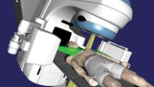
8) Proton Radiation. While most radiation beams harness their power from electrons, clinicians have started using protons to treat lung tumors. Because positively charged, subatomic protons only travel a limited distance through the body, proton radiation is thought to cause less damage to surrounding healthy tissue when compared to X-rays traditionally used in radiation therapy.
Proton-based radiation therapy is particularly useful for 4-D adaptive radiotherapy in treating lung cancer, where breathing can move the lung, making it difficult to hit the target. One obstacle to widespread adoption of proton radiation, however, is the need for an algorithm that quickly calculates the proton beam’s direction and intensity for each breathing phase.
9) Ambulatory Imaging. Picture a digital imaging detector built into a stretcher in an ambulance. In the few critical moments before reaching the hospital emergency personnel take X-rays of the patient. The images, beamed wirelessly to the hospital, enable the emergency room staff to begin treatment immediately upon arrival. Using organic electronics, scientists are expanding flexibility in the capture of X-rays. Such technology may be embedded in a hospital bed, a wheelchair, or even a hospital gown, enabling X-rays to be taken with minimal inconvenience to the patient. In addition, such devices free up hospital resources by eliminating the need to transport patients.
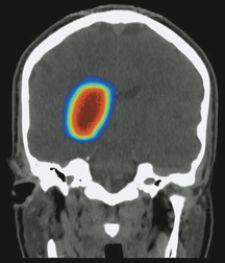
10) 3-D Surgery. Surgeons have traditionally relied on 2-D X-rays to view affected areas before surgery, and on invasive techniques to locate hard-to-reach tumors or damaged organs once in the operating room. Image-guided surgery uses CT scans, MRI and light emitting diode cameras to create 3-D real-time images of the surgical field and the precise location of tumors.
Image-guided surgery has many applications and is particularly useful to neurosurgeons in their preoperative assessments for brain tumor surgery where surgeons can more confidently remove brain tumors without causing new injuries to delicate structures.

 July 16, 2026


