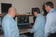
Physicians at the the Illinois Neurological Institute work together to create a treatment plan.
The name of the game in radiosurgery and radiotherapy is delivering the highest possible dose to the target volume and sparing the surrounding normal tissue and the structures. That requires immobilizing the target and drawing up a good plan, according to E. Ishmael Parsai, Ph.D., chief of Medical Physics, Medical College of Ohio, Toledo. The key to creating an individualized plan for accurate dose delivery for each patient goes beyond sophisticated software — it takes teamwork.
The radiation oncology treatment team is comprised of a combination of specialized medical professionals, including a radiation oncologist, neurosurgeon, medical radiation physicist, dosimetrist, radiation therapist, radiation therapy nurse and neurologist or neuro-oncologist. While the radiation oncologist and neurosurgeon oversee treatment and interpret the results of these procedures, every member of the team contributes to process.
Teamwork Makes a Difference
A group effort can make all of the difference when challenged with a complex situation in radiation therapy.
The medical team the University of Pittsburgh Medical Center (UPMC) Cancer Centers had to specially design the treatment for a lung cancer patient using a new technique that combines image-guided radiotherapy (IGRT) with respiratory gating to zero in on her tumor while adapting for breathing motion. The clinicians successfully executed the treatment, while protecting the patient’s heart and esophagus as well as the healthy parts of her lung by delivering dynamic IGRT treatment using Varian’s Trilogy linear accelerator equipped with Varian’s On-Board Imager for monitoring tumor motion and the RPM respiratory gating system, which synchronizes beam delivery with the patient’s natural breathing cycle.
“We had to be concerned about compromising this patient’s ability to breathe,” said Dwight Heron, M.D., vice chairman of clinical affairs, department of Radiation Oncology, UPMC Cancer Centers. “As a result, it was vitally important to minimize the exposure of radiation to the surrounding healthy lung tissue.”
As the patient had to be repositioned each day, UPMC’s clinical team used the On-Board Imager to generate high-quality radiographic X-ray images of the targeted area and also produced 3-D cone-beam CT images to verify patient positioning. An imaging study revealed that the tumor moved 1.2 centimeters with every breath. This degree of tumor motion would usually result in extending the treatment margin by about 1 to 1.5 centimeters around the tumor. However, the clinicians used Varian’s RPM respiratory gating solution to limit the radiated area.
“We set our RPM respiratory gating system to deliver the treatment beam only during a particular part of her respiratory cycle, when her tumor moved the least,” Heron said. “This happened when she was about halfway through her exhalation – the tumor was virtually motionless at that point, so we could target it very accurately. Using this approach, we were able to decrease the treatment margin to half a centimeter. Technologies like the Trilogy machine, with the On-Board Imager and respiratory gating, make it possible for us to adopt a truly personalized approach to cancer care.”
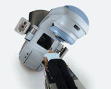
Effective SRS Management
At the Illinois Neurological Institute, a mid-state facility where patients travel 75-125 miles from rural communities, they treat metastatic brain and spinal column tumors on Varian’s Trilogy. Recently, they upgraded Trilogy with a mini-multileaf collimator (2.5 mm) designed for very small targets.
“The ability to use IMRT planning with the multileaf collimator and multiple beams allows for exquisite planning, avoiding dose to brain stem, spinal cord, and pharynx, esophagus, and salivary glands, significantly reducing the risk of morbidity and again often allowing the effectiveness and efficiency of a single SRS fraction. The infrared optical tracking system has been very helpful in alignment and tracking in frameless treatments in the brain and cervical spinal cord.
While the equipment is state-of-the-art at the clinic, the execution would be ineffective without the clinical team. Patrick Elwood, M.D., a neurosurgeon, sees the patients with James McGee, M.D., or Brian Griffin, M.D., both radiation oncologists. “We try to arrange for the CT simulation planning and a possible MRI for fusion that day so that one of the three physicists can begin the fusion and the planning process,” explained Dr. Elwood. “Most patients are treated either in a single fraction, or if because of anatomical dose limitations, five fractions.”
“I believe we are in the very early development of body radiosurgery and this will change very rapidly as the oncology community as a whole becomes aware of the new possibilities of effective management,” said Dr. Elwood. “I would expect this to change much more rapidly than the development of intracranial radiosurgery because it is getting started from a much wider database.”
Retrofitted for Accuracy
In fractionated stereotactic radiosurgery (FSR), neurosurgeons and radiation oncologists are first looking for accuracy in positioning the tumor and delivering the dose to sub millimeter targets, requiring more accuracy than a surgical knife.
At Medical College of Ohio in Toledo, where physicians have performed stereotactic radiosurgery since 1994, engineers recently retrofitted new equipment and a miniature multileaf collimator system by 3D Line Medical Systems to the head of the electrolinear accelerator. The miniature 3 mm-thick multileaf system includes a software that allows for inverse planning using IMRT.
“The 3D Line system uses aperture optimization through full arc, which is a more advanced technique than conventional weight optimization using IMRT,” said Dr. Parsai. “At each gantry angle as the beam is rotated around the head of the patient, the target is seen by the system and the miniature multileaf adjusts the target for optimum dose to that volume and sparing of the tissue that might be in the path of the beam.”
Once the critical structures are identified, the physicist generates the planning. This inverse planning method allows the physicians to decide what maximum radiation they can allow for any of the identified critical structures that might be in the path of the beam, entrance or exit alike.
Mapping Out a Plan
To create the ideal plan, clinical teamwork is probably the most important factor says Parsai. At Medical College of Ohio, a patient, for example, comes in at 7:30 in the morning. Neurosurgeons, radiation oncologists and physicists are all in the surgical suite where they attach the frame to the head of the patient after reviewing the MRI or CT of the patient, identifying exactly the target areas within the brain. This is done on a consultation basis. The neurosurgeon then puts on the frame to the head of patient after they acquire new images – CT or MR or both or sometimes PET – with the frame attached to the head of the patient.
Then after all of these are done the images are sent to a computer where all members of the team will gather around and review carefully, slice by slice, the current status of that patient and make the decision of the best way to approach.
“Neurosurgeons, oncologists and physicists are sitting there reviewing the slices of images and deciding what is the best way to approach. So even though each individual has done some preliminary work on their own, when you put a few people in a room, then their ideas on the best approach is considered. Each person presents their own expertise and I think a decision is made,” indicated Dr. Parsai.
“For physicians, neurosurgeons, and radiation oncologists, their expertise is to know normal versus abnormal tissue structures. Everyone respects the others’ expertise. There might be discussions on what they include or exclude for a target volume. The importance of tiny areas margined with a tumor into the target as is related to the quality of life of the patient or survival of the patient are really reviewed very carefully. Neurosurgeons are given the primary decision-making on delineating the target and delineating the structures that could be damaged as a result of significant radiation. Radiation oncologists also have the same expertise and knowledge,” said Dr. Parsai.
Once the neurosurgeon and radiation oncologist delineate the target volume as well as the critical structures and they identify the maximum dose tolerance to the critical structures, the radiation oncologist will write a prescription dose to the target volume. Then the physicist will take over the process of developing a treatment plan to best meet the requirements set forth by the radiation oncologist.
This team approach continues until the plan is reviewed and approved by the radiation oncologist.
“After the plan is generated by the physicist,” said Dr. Parsai, “in the evaluation and process, the plan is often changed based on the discussions with radiation oncologists and various optimizations that are presented to result in a better solution.”
The patient is brought down after that and quality assurance (QA) is performed on the patient to verify that the position automatically provided by the system exactly matches the intended target. Those are approved and verified through portal images. Then the dose is delivered and often these patients are sent home that same evening.
“A lot of education goes on in these rooms,” noted Dr. Parsai. “It is a very unique situation.”