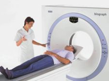
Siemens Biograph PET/CT series consists of two-, six-, 16-, 40- and 64-slice configurations and provides high-imaging quality and clarity.
Myocardial perfusion imaging (MPI) using single photon emission computed tomography/computed tomography (SPECT/CT) is an application that has been recently bolstered by the development of quantitative tools that enhance the system’s clinical capabilities in the nuclear cardiology community.
Similarly, the use of positron emission tomography fused with computed tomography (PET/CT) is well established in oncology for diagnosis and staging, however, there is a trend among clinicians toward using PET/CT for cardiac imaging, levering its superior image quality, visualization and quantitative capabilities. Yet the adoption of PET/CT for MPI is notably slow due to several contributing factors.
Pros, cons of cardiac PET/CT vs. SPECT/CT
Over the last year, vendors have introduced a series of new hybrid SPECT/CT systems and software solutions, including GE Healthcare’s Volumetrix Suite with automatic registration and 3D image rendering, which expedites patient throughput and streamlines workflow. Another is Siemens’ SMARTZOOM collimator, which is referred to as a cardio-centric orbit that enables the heart to remain within the collimator’s magnification area throughout the entire study, enabling the study to be performed in less than one minute. Adding to the list is Philip’s Brightview XCT system, which offers the only scalable hybrid camera to fit into a 12-by-15.5-foot room, thus requiring little to no room renovation and reducing the overall cost of ownership.
Despite the growing number of SPECT/CT tools with enhanced capabilities dramatically improving image quality, the current generation of PET/CT systems still produces superior images, according to Partha Ghosh, M.D., clinical marketing manager, molecular imaging, Siemens Medical Solutions USA.
“Cardiac PET/CT uses a short-acting isotope and higher doses can be injected. This, when combined with the higher sensitivity of PET, contributes to the overall improved image quality,” said Dr. Ghosh. “Thus, in obese patients or in patients who have thick chest walls or women with large breasts, we expect PET/CT to be definitely superior. Other advantages of PET/CT include the use of short-acting isotopes, which are true perfusion agents. Due to this, both stress and rest imaging can be performed in a relatively short time. This is a major advantage over SPECT/CT, requiring about two to three hours between stress and rest, which in turn, increases the total procedure time. We can perform blood flow quantifications much more effectively and reproducibly with PET/CT due to dynamic imaging capabilities. Thus with PET, we have better image quality, better visualization in obese patients and better quantitative capability.”
PET/CT is also capable of absolute quantification of myocardial blood flow and flow reserve estimation. This helps overall visual interpretation of myocardial perfusion by providing flow reserve estimations, which provide higher diagnostic confidence, particularly in situations where there are borderline changes and also in situations with balanced disease. In balanced disease, all four vessels are equally diseased, in which case there is a generalized decrease in myocardial blood flow. Due to the global reduction in blood flow, the perfusion images can artificially appear to be normal. PET/CT is able to accurately identify areas of abnormality in balanced disease, according to Dr. Ghosh.
Despite the value of this capability, it is still not widespread and is used only in certain major research centers. “However, we are working to make this process user-friendly so that people can use it on a regular basis. Initial calculation methods require input from physicists, but we are trying to make it simple so that standard users can use it more effectively.” Dr. Ghosh said.
New agent extends shelf life
Another factor weighing in on the advantages of PET/CT over SPECT/CT is the development of new agents for cardiac PET/CT. Bracco Diagnostics recently introduced an agent for cardiac PET/CT perfusion imaging - CardioGen-82, a Rubidium Rb 82 generator, which is currently the only generator-based PET/CT agent reimbursed for the evaluation of coronary artery disease. The physical half-life for Rb-82 is 75 seconds, which allows for an efficient protocol. By the time the imaging is completed for the rest portion (approximately six minutes), stress can follow immediately and then the stress images can be acquired (approximately six minutes). The study can be completed in approximately 40-45 minutes. Key features of Rb-82 are that it has properties similar to Thallium-201 (Tl-201), defects are visualized within two to seven minutes after injection, and due to its short half-life, both sets of images can be acquired using the same sized dose.
Slow to catch on
With all of the advances in PET/CT perfusion imaging, why isn’t it more widely adopted for cardiac imaging? According to Dr. Ghosh, the main impairment to its widespread adoption is the limited availability and expense of Rubidium, which is the main isotope required for PET/CT perfusion imaging. Another factor is the limited expertise in cardiac PET/CT among nuclear medicine physicians.
However, Dr. Ghosh noted, “We expect this to significantly improve in the next few years because we are expecting new radiopharmaceuticals — Fluorine-18 labeled myocardial perfusion agents — which will have a much better logistic advantage for cardiac PET. In the next two to three years, we hope to have this new radiopharmaceutical available, and we will be able to see much more utilization of PET/CT in cardiology. There is also a significant need for us to educate the general PET/CT users on the value of PET/CT in cardiac imaging, although I see more and more physicians are getting interested.”
Others attribute PET/CT’s slow penetration in cardiac imaging to a lack of coverage for FDG-PET/CT in non-oncology applications. According to Frost and Sullivan research analyst, Travis Chong, this has placed a cap on near-term market growth and left it susceptible to disruptive technologies from competing modalities in the long-term future. Chong notes that data released by the National Oncology PET/CT Registry (NOPR) in March 2008 revealed that FDG-PET/CT scans result had a 36.5 percent change in the decision of whether, or how to, treat a patient’s cancer in approximately 23,000 patients. “As more studies clearly demonstrate the positive clinical impact of molecular imaging, advocates of the modality again question whether the CMS’ requirement for “Coverage with Evidence Development” is the best strategy for coverage expansion,” he said.
Other hindrances to using PET/CT for cardiovascular imaging could be the average cost of a system at around $1.6 to $2.1 million. One strategy to overcoming economic obstacles is to cut the system in half. Positron Corp. looks to strengthen PET/CT perfusion’s position with its recent introduction of a PET-only dedicated cardiac camera.
“Currently under development, this ‘economically priced’ PET system is speculated to cost approximately $600,000 dollars. With a small footprint and lower costs, the PET camera is expected to directly compete with the mobile dedicated-cardiac SPECT/CT cameras within the primary-care physician and cardiologist office setting, which represents the largest (and most lucrative) untapped market segment within the MPI industry, said Chong.
With the wave of new radiopharmaceuticals being developed for cardiac imaging, the development of more affordable PET/CT systems specifically designed for cardiac use, the recent attainment of coverage for the Rubidium-82 generator and growing interest among its users, PET/CT in the MPI market is well on its way to attaining its place as the prime choice for cardiac imaging. “If PET/CT perfusion is able to successfully penetrate this market segment,” said Chong, “the modality may finally be able to shake off its misconception of being an ‘oncology-only’ modality.”