Accuray's robotic radiosurgical device, the CyberKnife System
In today’s world, it’s hard to understand that a regimen of surgical resection followed by radiation and chemotherapy – a procedure practiced in medicine for over 100 years – is still the standard of care in the treatment of breast cancer.
But amazing advancements may one day obviate the need for surgery at all thanks in part to emerging technologies such as cryoablation and radiofrequency heat and laser ablation – technologies that are not only catching cancer cells earlier, but obliterating them without the damaging effects of radiation and chemicals.
Many current advancements in breast tumor treatment are made possible by remarkable improvements in imaging, such as digital mammography, ultrasound and MRI, which have allowed radiologists to spot tumors in their infancy when they can be removed or treated by technologies other than radiation.
Current Landscape
The National Cancer Institute identifies the four types of “standard” treatment for breast cancer today are surgery, radiation (either whole or partial-breast), chemotherapy and hormone therapy. Breast conservation therapy (including lumpectomy, partial mastectomy and lymph node dissection), in which the tumor and a margin of surrounding normal tissues are surgically removed, is increasingly being used for patients with early-stage cancers. Close to 80 percent of patients can be successfully treated today with breast conservation therapy, according to the Radiological Society of North America.
Even breast conservation therapy, however, has gradually come into disfavor with many women and radiologists; the chances of cancer recurrence still looms, and the procedures are often disfiguring and painful.
The Cancer Institute also notes that newer types of treatments such as sentinel lymph node biopsies (a cancer staging technique), high-dose chemotherapy with stem cell transplants and monoclonal antibodies as adjuvant therapy are showing a great deal of promise. “There are a number of compounds that target specific cell receptors and thereby provide more targeted treatment than conventional chemotherapy agents,” said Susan Levine, Ph.D., vice president of Technology Assessment, Lansdale, PA-based Hayes Inc., a technology assessment firm.
Still, conventional tumor treatments have been known to miss cancer cells in the tissue margins surrounding the cavity where the removed tumor once resided, resulting in additional radiation and/or chemotherapy. As many as 55 percent of breast cancers removed by lumpectomy, for example, have been found to leave cancer cells behind, according to published reports. In addition, conventional treatments can often be hampered by challenges in immobilizing breast tissue.
Significant advancements in surgical excision, however, are now addressing those concerns.
Aliso Viejo, CA-based SenoRx Inc. is exploring the development of radiofrequency tools for breast surgical excision, according to Doug Bradley, director of Marketing. While the company’s radiosurgical scalpel, ShapeSelect, is not yet approved for breast cancer surgery, SenoRx is investigating future RF excision applications, Bradley said. The company’s patented Anchor Guide fixation system is used extensively in brachytherapy procedures, Bradley said.
TeraView Ltd., Cambridge, England, meanwhile, is exploring the use of “terahertz light,” which falls between microwaves and infrared light, in guided surgery for breast cancer. CEO Don Arnone said TeraView’s technology, called terahertz pulsed imaging (TPI), has been able to spot cancer below the epithelium, where about 15 percent of breast cancers have been known to originate. Arnone said TPI is non-ionizing and less hazardous than conventional X-rays. “Our technology is likely to be used mainly for diagnostic purposes, as well as in guided surgery,” he said. “The ability to intra-operatively image tissue and determine the location of cancerous and potentially precancerous lesions will result in better patient outcomes.”
Brachytherapy’s Push
High-dose breast brachytherapy is showing considerable promise not only as a viable alternative to mastectomy, but may one day rival whole breast external irradiation in the fight against early stage breast cancer. Also called accelerated partial breast irradiation, the procedure brings the radiation inside the cavity left from a lumpectomy in the form of tiny “seeds” made of titanium or other material and encapsulating a naturally occurring isotope, such as iridium 192.
High-dose breast brachytherapy typically entails 10 treatments (also called “fractions”) over a period of five days. During each treatment, performed twice a day, the seeds are placed into the lumpectomy cavity by special wires inserted through tiny catheters, left for a small, predetermined amount of time, and then removed. The number of seeds, the dosage and placement of each seed are determined by special treatment planning software. High-dose brachytherapy was most commonly administered through a technique called “interstitial high-dose rate multicatheter brachytherapy,” in which up to 30 catheters were inserted into the breast and left there during the five-day treatment regimen.
In 2002, Proxima improved on the technique with a procedure called “intracavitary balloon catheter brachytherapy” (trademarked as “MammoSite”), in which a deflated balloon was inserted into the lesion bed and inflated with saline. Instead of multiple catheters at various locations through the breast tissue, only one catheter was needed. Today, balloon brachytherapy is the most widely practiced form of high-dose breast brachytherapy, chiefly because it is simpler to perform and as effective as its predecessor.
High-dose brachytherapy has many advantages over whole breast radiation, including fewer side effects, decreased hospitalization, reduced risk of postoperative infections and faster transition to chemotherapy because of shortened treatment times. Fremont, CA-based XOFT Inc., recently received Food and Drug Administration marketing clearance for a seed alternative, a device best described as a tiny X-ray tube.
In October, Aliso Viejo, CA-based BioLucent received FDA clearance to market its SAVI applicator, a multicatheter, single entry approach to breast brachytherapy. The SAVI essentially combines the features of the interstitial technique with those of the MammoSite technology. “Our device is a hybrid of both techniques,” noted Jill Anderson, BioLucent president. The physician expands the catheter bundle by turning a mechanism from outside the breast. The catheters expand to form an ellipsoidal shape inside the cavity. Delivery of radiation through the device’s individual catheters allows the doctor to better contour and control the radiation dose.
“The SAVI essentially gives control back to the radiation oncologist by allowing him to shape the dose,” Anderson said. “Each channel can be turned on or off, which allows more precise delivery of radiation to the margin surrounding the tumor bed.”
Emerging Technologies
In recent years, there has been extensive exploration into applying proven technologies to breast tumor treatment, particularly in early stage cancer and small breast tumors. Many of them are nonsurgical and can be performed on an outpatient basis by interventional radiologists.
Such emerging techniques include photodynamic therapy, energy-based ablation, such as radiofrequency and focused ultrasound and microwave thermotherapy, and other ablation procedures, such as laser ablation and cryoablation. These techniques use alternative forms of energy to achieve complete cell death.
Ablation techniques are showing a great deal of promise, and offer such benefits as: faster healing; noninvasive or minimally invasive; shortened treatment times; improved quality of life; greater dose precision; breast preservation; possible avoidance of additional follow-up surgery, radiation therapy or chemotherapy; and lower costs (from avoidance of surgery and overnight stays). Other techniques include the following:
Radiofrequency ablation (RFA) – A well-established technology that has been used successfully to stop bleeding, remove bone growths and skin lesions, RFA also has shown promise in treating liver cancer and heart rhythm disorders. RFA employs the use of small probes, which are inserted through the skin and directly into the tumor. An umbrella-like series of prongs then extends outward, delivering an electrical current into the tissue, causing friction and with temperatures approaching 200 degrees Fahrenheit, “cooks” the tumor. RFA has a litany of benefits. It has been shown to reliably kill a small targeted volume without damaging healthy tissue, is accurate and easy to control, possibly avoids the necessity of re-excision, has a very low complication rate, is minimally painful, results in low blood loss and is safer because the energy is absorbed into surrounding tissues as simple heat.
Established players include RITA Medical Systems and ValleyLab, although both companies currently are not marketing their devices specifically for breast tumor treatment.
One variation on thermal radiation techniques, such as RFA, has been developed by privately held Oncobionic Inc., which claims its irreversible electroporation (IRE) technology safely treats a wide range of diseases from liver tumors to prostate cancer. IRE technology uses needles and image guidance similar to existing thermal ablation technologies, but instead of “cooking or freezing” the targeted tissue, IRE disrupts the cell membrane, destroying the targeted cells without thermal damage and without affecting connective tissue and structures, such as blood vessels and ducts. In late October, Queensbury, NY-based AngioDynamics entered the field with its $25 million acquisition of Oncobionic. Marketing of the technology is slated to begin in mid-2008.
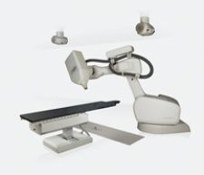
Sunnyvale, CA-based Accuray Inc., is currently exploring the use of its robotic radiosurgical device, the CyberKnife System, as a tool for ablating breast tumors. The device delivers multiple beams of precisely directed radiation that converge upon a tumor, minimizing damage to surrounding healthy tissue. In contrast to traditional radiation therapy, robotic radiosurgery combines image guidance technology and computer controlled robotics, which enable the system to deliver high doses of radiation with sub-millimeter accuracy, according to Kristine Gagliardi, vice president of Clinical Development.
Accuray received FDA clearance for treating tumors anywhere in the body in 2001 and a breast treatment protocol is in the early stages of development at a number of academic medical centers. Some of the challenges to work through are the dosing regimen, immobilization of the breast and tracking during treatment, Gagliardi indicated. Joanne Davis, clinical program manager, added that the CyberKnife System may be used as a radiosurgical tool following lumpectomy.
Laser ablation – This technique is said to heat and destroy breast tumors through the use of an imaging-guided fiberoptic cable placed into the tumor. A quartz at the tip of the cable emits laser light energy, destroying a small tumor and at least half a centimeter of margin tissue. Clinical trials with laser ablation have been moderately successful; some cancer cells have remained after the procedure and subsequent surgical resection, according to published reports.
Focused ultrasound therapy – A noninvasive thermal ablation technique, imaging-guided focused ultrasound therapy directs energy waves through several transducers on the surface of an immobilized breast, resulting in temperatures as high as 70 degrees Celsius. The technique reportedly has been used in a pilot study to treat patients with inoperable breast cancer.
Focused microwave therapy – Reportedly based on the Reagan Administration’s Strategic Defense Initiative or “Star Wars,” technology (which was developed in the 1980s to seek and destroy nuclear missiles), focused microwave therapy (FMT) exploits the difference in water content between normal cells and cancer cells. Like focused ultrasound, this technology is noninvasive. MIT developed the technology for cancer treatment and licensed its use for cancer treatment with Columbia, MD-based Celsion Corp. Other companies, such as ValleyLab, also are exploring the use of FMT for breast tumor treatment.
Cryoablation – Like RFA, cryoablation has long been used in medicine to treat other diseases such as liver and prostate cancer, and conditions such as skin lesions. Cryoablation was recently cleared by the FDA to treat benign breast fibroadenomas. The procedure has only shown effectiveness in treating early stage breast cancer, and very small tumors less than 1.5 cm in size. Cryoablation employs the use of a tiny, thin probe, which is inserted directly into the tumor. The tip of the probe emits freezing temperatures, shuts off to allow the tissue to thaw, and repeats the process several times. Through the use of substances such as argon gas, an ice ball surrounds the necrotic tissue. Scientists claim that the body safely resorbs the dead tissue, usually within six months.
The procedure requires no sedation or anesthetic (freezing numbs the tissue), and there is reported evidence that the process actually stimulates the immune system to destroy cancer cells elsewhere in the body. In addition, the resulting ice ball is said to be visible with sonography in real time, giving greater control and assurance that the tumor has been destroyed. Published reports also state that overall costs for cryoablation could actually be lower than those for a lumpectomy and similar to those of a biopsy. Pleasanton, CA-based Sanarus Medical Inc. currently markets its Visica Treatment System as an office-based cryoablation treatment for breast tumors.