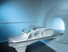
The Aurora System with Bilateral 3-D SpiralRODEO is an FDA cleared, fully integrated MRI system designed specifically for breast imaging.
The radiation oncology community is increasingly more optimistic about winning the battle against breast cancer. This hope is in part due to the many developments in magnetic resonance imaging (MRI) for breast cancer used for diagnosis, staging, pretreatment and preoperative planning. Simultaneously, researchers are developing new techniques in radiosurgery for treating cancerous tumors in the breast and are turning out promising results from these preliminary studies.
Much discussion regarding MRI for breast diagnosis has centered around high false positive rates and the expense. However, recent studies demonstrate the value of MRI for breast in optimizing treatment. A recent study at the University of Florida found that MRI used for preoperative/pretreatment planning for breast cancer reveals important diagnostic information that changes the treatment plan about 20 percent of the time. In this study, 79 women who had localized noninvasive or early stage invasive breast cancer and who were planning to have a lumpectomy underwent MRI for preoperative evaluation, in which case MRI-guided biopsies revealed additional cancer about 40 percent of the time. Ultimately, approximately 75 percent of patients underwent the planned upon lumpectomy, while one-fourth had a total mastectomy.
Elsie Levin, M.D., radiologist and director of the Faulkner-Sagoff Breast Imaging and Diagnostic Centre, one of the early adopters of breast MRI, has found similar results from her practice using the Aurora 1.5T Dedicated Breast MRI System for pretreatment and preoperative planning due to its ability to provide important diagnostic information. The Aurora System with Bilateral 3-D SpiralRODEO is an FDA cleared, fully integrated MRI system designed specifically for breast imaging. “If you look at the literature in general, in the newly diagnosed cancer patients, MR will show additional disease in about one-third of patients and about one-half of those patients it actually changes surgical planning so that’s about 15 percent,” indicated Dr. Levin.
Some controversy surrounding the increase in MRI use for breast may mean more unnecessary mastectomies. However, Elizabeth Jekot, M.D., a dedicated breast radiologist and founder/director of The Jekot Breast Imaging Center in Richmond, TX, also an Aurora user, pointed out, “Now when we say alter treatment, it doesn’t mean that we went from a lumpectomy to a full mastectomy. It might mean that they biopsied an additional area to show the extent of margins. It might have changed it minimally. I think that there is this fear that if we do all of these MRIs, we might be converting all of these lumpectomies to mastectomies, and I
don’t think that’s true. I think that what we’re doing is actually confirming the extent of disease in some patients who may need mastectomies, but at the same time reassuring patients who have had small cancers that a lumpectomy is an appropriate surgery for them. With that, I think that we’re decreasing the number of failed lumpectomies.”
Map out treatment with MRI
With breast cancer patients at a 50 percent rate of reincision after their original surgery, according to Dr. Levin, MRI planning is crucial. MRI optimizes treatment by enabling cleaner margins in surgery and locating all disease in the breast(s). Although most cancer recurs in the location of the original tumor, five percent of the time the cancer recurs in a completely separate quadrant of the breast. When considering partial breast radiation therapy, it is paramount to clean margins and find all cancer.
“You’re only going to be treating that tumor bed, so I think particularly in that situation where you’re not treating the whole breast and where margin assessment is very important, it’s nice to know ahead of time really what you’re dealing with, whether or not they’re going to be an appropriate candidate for that therapy,” Dr. Levin said.
Dr. Jekot concurred, saying, “It’s not only helpful with the ipsilateral breast but also the contralateral breast insofar as making sure that there is nothing there that needs to be addressed at this time. We want to make sure that we’re not overestimating or underestimating the extent of disease so that she can basically optimize her preoperative planning so that she can get the definitive surgery from the get go. So, in addition to that, it gives us, particular to the Aurora, a very good look at the lymph node area or axilla insofar as determining whether or not there might be some pathologic lymph nodes.”
Dr. Levin also stressed the clinical importance of scanning both breasts with MRI after a diagnosis has been made in one breast. She explained, “If you scan both breasts which you should do, the chances of finding a cancer in the opposite breast, which has been found to be normal after ultrasound and physical exam, is anywhere from three to nine percent depending on the study.”
MRI is also a valuable diagnostic tool in terms of its ability to detect lobular cancer, which is difficult to detect on mammography, determining whether or not the tumor has penetrated the chest wall muscle. Recent studies have also demonstrated MRI’s higher sensitivity in detecting ductal carcinoma in situ (DCIS) particularly of the high-grade type, compared to mammography, noted Dr. Levin. Locating all possible sites of cancer and accurate margin assessment is crucial in planning for
surgery and treatment.
Reimbursement for BMRI
Despite the effectiveness of breast MRI (BMRI) for preoperative and pretreatment planning, clinical utility depends on whether there is reimbursement.
According to Aurora Imaging Technology, Inc., the Centers for Medicare and Medicaid (CMS) 2008 Final Rule for the Hospital Outpatient Prospective Payment System allowed reimbursement for guidance services, including MR guidance, image processing services, intra-operative services, imaging supervision and interpretation services, diagnostic pharmaceuticals, contrast agents and observation services. Specifically, reimbursement for BMRI with contrast agent for breast and bilateral images are up 6 percent to $397.13 and for MRI without followed by with contrast for breast and bilateral images are up 5 percent to $525.24. The decision by CMS to reimburse for BMRI procedures is testimony to the success of the technology.
Radiosurgery shrinks
tumor margins
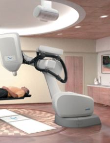
Also promising are results from new radiosurgical techniques that carve out sharper, tighter edges around cancerous tumors. Clinicians are shrinking the margins of healthy surrounding tissue performing radiosurgery with technology such as Accuray’s CyberKnife Robotic Radiosurgery System, a robotic radiosurgery system designed to treat tumors anywhere in the body noninvasively and with submillimeter accuracy.
Sandra Vermeulen, M.D., radiation oncologist and co-director of Seattle Cyberknife uses the Cyberknife on an experimental basis for the treatment of breast cancer. The National Surgical Adjuvant Breast Project has been conducting a clinical trial for nearly three years involving partial radiation therapy versus whole radiation for breast cancer. Although the trial is not yet closed, the data thus far looks favorable for being able to treat the small area of the breast at the site of cancer and obtain the same favorable outcome as if a woman had a mastectomy, according to Dr. Vermeulen. Due to the data from this trial and the release of cyberknife technology, which tracks motion while radiation is administered, Seattle Cyberknife began investigating the use of cyberknife in the treatment for breast cancer in a motion tracking study.
“What we do know in theory is we should be able to reduce the volume around the lumpectomy cavity after the surgeon takes the breast cancer out compared to what we’re looking at right now using conventional machines, which is putting a 27 millimeter margin around the target and treating it for five days,” said Dr. Vermeulen. “And now we can reduce it to 10 millimeters and get an even better result. So it’s even less normal tissue and less side effects, and so that is what everyone’s gotten very excited about.” The hope is that reducing dose delivery to healthy tissue will result in fewer side effects from radiation treatment and a better prognosis.
Another new radiotherapy device used for many types of cancer including breast is Varian Medical Systems’ RapidArc for faster image-guided intensity-modulated radiotherapy (IMRT). According to Varian, RapidArc delivery produces as little as 600 MU per fraction compared to fixed-field IMRT treatments that yield 1,000 to 3,000 MU. This new technology consists of a single gantry rotation and speeds treatment delivery by two to eight times faster than conventional IMRT, which may also result in less scatter, less leakage and lower doses to peripheral areas. Ultimately, this system may lead to shortened treatment times and increase the accuracy of tumor targeting. Specifically to breast cancer, the exceptional tumor targeting will reduce the risk of exposing the heart and lungs to radiation.
With new realizations in the use of MRI for breast in preoperative and pretreatment planning and significant advances in breast cancer treatment itself, doctors are gaining in the war on breast cancer, one battle at a time.