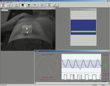
Varian's RPM respiratory gating system alerts the physician when there is any movement.
While many professionals in the radiation oncology field insist that true adaptive radiation therapy is not yet taking place due to a lack of time and resources, a growing number of these professionals are in fact “adapting” technology to make it suitable for treating a wide array of cases with more precision and accuracy.
Of utmost importance to radiotherapy treatment providers is to treat the tumor and spare the normal tissue. The limitations that impede delivering treatment accurately and efficiently are tumor motion and visualization. However, many radiation oncology professionals are discovering a variety of ways to better gauge that motion and obtain clearer, more accurate images and, in the end, more effectively treat the tumor.
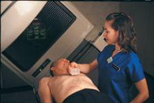
Respiratory motion is one of the main sources of anatomical motion. The approach taken by Jerome Landry, M.D., chief of Service at Grady Memorial Hospital in Atlanta, GA, is to “evaluate the phases of respiration when tumor motion is minimized and allow treatment during this particular phase.” Dr. Landry relies on Varian’s respiratory gating system, RPM, to assist in accurately targeting the tumor.
Before Todd Scarbrough, M.D., medical director of the MIMA cancer center in Melbourne, FL, used Varian’s RPM respiratory gating system to estimate motion, like most physicians, he had to use larger margins. Dr. Scarbrough explains that when regulating tumor motion without the respiratory gating system, 1.5 cubic cm are added to the treatment volume in contrast to only a 5 cubic mm margin when using RPM. In other words, when treating a 3 cubic cm spherical tumor without using RPM, an additional 15 cubic cm of normal tissue is damaged.
But does this really make a difference for patients? Dr. Scarbrough explored this exact question and recently released the results of a study, which demonstrates the benefits for patients when using Varian’s RPM respiratory gating system. In his study, patients who were simulated using gating or a deep inspiratory breath holding (DIBH) technique showed a two-year overall survival of 100 percent when compared to 66 percent for those treated using free breathing (FB), the more conventional approach. In addition, freedom from progression for a two-year interval was also 100 percent for those using respiratory gating or DIBH and only 22 percent for FB patients.
While controlling for respiratory motion with gating positively impacts treatment outcomes, there are still many other types of motion that could negatively impact treatment if not considered. To this end, the FDA recently gave 510(k) clearance for Varian’s patient position monitoring capabilities that have been added to its RPM respiratory gating system to synchronize imaging and radiation therapy treatment with a patient’s respiratory cycle. The new feature reportedly detects any motion that compromises the accuracy of the treatment, and works by placing a reflective marker box on the patient’s chest or abdomen and monitoring its motion using special cameras positioned in the treatment room. The new version of the RPM system analyzes the marker block motion in all three dimensions as it moves up and down, forward and back, and side to side. A new display on the system’s console immediately alerts the radiation therapist if the patient moves and should be repositioned.
When asked about the impact of this new technology on patient treatment, Dr. Scarbrough stated, “It will depend on how much moving of the patient that you’ve got. But everything in radiation is getting to the point that what you don’t know is going to hurt you. It’ll be used, but how useful will it be? Well, let’s define useful. If it corrects one out of 100, then it may be.”
Another tool improving treatment is the TomoTherapy Hi-Art System, which leverages the ring gantry geometry used in CT scanning for the delivery of state-of-the art, intensity modulated radiation therapy (IMRT) from all angles around the patient. According to the Tim Holmes, Ph.D., co-creator of tomotherapy, “I think the most important aspect of tomotherapy is the high level of integration in the design of the device. An obvious example is the integration of the treatment planning and delivery systems on a common database. Conventional radiotherapy systems typically have two or more databases – one for the treatment planning and one for the treatment delivery and possibly another for imaging. This high level of integration allows one to develop new procedures that utilize the integrated CT imaging, like CT simulation and treatment using the tomotherapy unit or adaptive radiotherapy.” With this type of integrated technology, tomotherapy is able to treat a wider array of cases more efficiently, accurately and precisely.
Holmes points to the clinical example of treating a lower limb Kaposi sarcoma. Due to the length and width of the treatment volume, it would be difficult to treat using a conventional radiotherapy system. However, TomoTherapy's unique features, including the high-resolution multileaf collimator and an integrated CT imaging system, allowed Holmes to treat the long volume as well as ensure that the thin shell of tissue was properly located in the beam irradiation path. Holmes stated, “It would be very difficult to achieve this type of dose distribution with a conventional radiotherapy machine due to the length of volume. One would also need to irradiate from many fixed beam directions using a conventional linac to approximate the dose distribution produced by rotational delivery.” According to the manufacturer, the treatment is easier to plan and deliver using tomotherapy.
In addition, the system offers an adaptive planning tool, in which one can pull up initial images of the patient on a workstation, overlay those images with the daily CT images, make adjustments and create a new plan. “It’s a very seamless process,” Dr. Holmes indicated. “It’s basically a re-simulation of the patient and more time efficient for physicians. It’s the poor man’s approach to 4-D imaging. Tomotherapy is consistent with what’s always been done. You modify volume to take account of motion when scanning. It’s really about getting a better estimate, not under-estimating and under-dosing the treatment.”
Another advantage is that in contrast to the helical CT, which uses kilovoltage, tomotherapy uses a megavolt CT (MVCT). “Kilovoltage produces nice images, but they are more prone to artifacts,” said Dr. Holmes. “Megavolt is clearly better because you don’t get metal artifacts or shadowing, which means that you get more accurate dose calculations.”
One system that may be the model for the next generation of radiosurgery units is the CyberKnife Robotic Radiosurgery System by Accuray Inc. By combining continuous image-guidance technology with a compact linear accelerator that has the flexibility to move in three dimensions according to the treatment plan, Cyberknife is designed to track, detect and correct for tumor and patient movement in real time, during the procedure, enabling delivery of precise, high dose radiation typically with sub-millimeter accuracy. One of the unique features of this unit is that “the CyberKnife linac is attached to a commercial robot to provide greater movement and achieve angles that fixed gantries cannot attain,” explained Iris Gibbs, M.D., co-director of CyberKnife Radiosurgery Program and assistant professor, Radiation Oncology, Stanford University.
Although technology continues to inch toward true real-time adaptive radiation therapy, until dose delivery reaches 100 percent accuracy, the procedure will continue to rely to a large degree on the finesse and skill of the radiation oncologist.