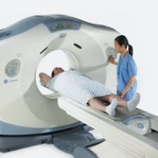
BrainLab's iPlan Flow software for brain tumors supports the planning of radiotherapy treatments in combination with local fluid delivery.
For years, intensity modulated radiation therapy (IMRT) has assisted doctors in contouring radiation beams around anatomical structures to target tumors. As tumors or organs shift, however, the beam can fall off target and damage surrounding healthy tissue. With image-guided radiation therapy (IGRT), which allows physicians to scan and locate the tumor while the patient is in the treatment position, the volume of healthy tissue exposed to radiation during treatment has shrunk from half an inch or more
to submillimeter sizes. IGRT also permits more powerful doses of radiation, and thus shorter treatment times and improved cure rates.
“I’d say IMRT has improved dosing of the target, and IGRT has improved targeting,” said Dwight Heron, M.D., director of Radiation Services for UPMC Cancer Centers. “The IGRT concept really is going to be the next great leap forward.”
Despite these advances, without improved targeting, healthy structures near tumors are still at great risk of injury. The new formulas for achieving greater conformality in radiation therapy add up to 4-D CT imaging and a technique known as six degrees of freedom (6-D) – tools that are accelerating radiation therapy and surgery into a more precise future.
4-D Tracks Moving Targets
Hitting a moving target may be the greatest challenge in radiation therapy today, and precisely localizing a tumor begins with imaging. When capturing a 3-D CT image of the patient, motion due to scan time and the relationship between the CT data acquisition period and the respiratory/cardiac cycle may produce significant artifacts. Motion artifacts often occur at the boundaries of anatomical structures, including target volumes and organs, making it difficult to delineate critical structures. This may cause erroneous positioning, shape and volume information for the target volume and other regions. Artifacts may also simulate disease, leading to further targeting misfires.
To correct artifacts produced by respiratory-induced motion of tumors and normal tissue, four-dimensional (4-D) techniques are used to achieve highly conformal radiotherapy during imaging, planning and radiation delivery. To quantify the respiratory cycle, a complete CT data set of images from each portion (bin) of the breathing cycle is obtained to produce a 4-D CT image – the challenge is to acquire it with minimal motion artifacts. Several methods may be used to acquire such a data set, including techniques that shorten scanner rotation times (fast scanning), prospective respiratory/cardiac gated image acquisition and retrospective image reconstruction. One of the greatest benefits of 4-D adaptive radiation therapy technology is that it facilitates a personalized approach to cancer care.
Sharpening IGRS Tools
As each person’s disease is unique, patients present individualized clinical challenges such as soft tissue deformation, positioning correction, tumor growth or shrinkage, irregular respiration and spontaneous motion. This is where 4-D is critical for adjusting to the natural movement of each patient.
Extracranial treatments for the spine, lung, prostate, liver and pancreas are on the rise in the U.S. Treating tumors in these areas is complicated by movement from the respiratory cycle. This has spawned the development of tools like the CyberKnife Robotic Radiosurgery System by Accuray, which provides integrated tracking systems, including the Synchrony Respiratory Tracking System for radiosurgery and Xsight, a spine tracking system for spinal tumor treatments. As image-guided technology locates the tumor, the system uses 4-D technology to track tumor position in real time, synchronizing radiation delivery to the motion of the tumor throughout the respiratory cycle.
Varian’s Clinac and Trilogy accelerators are also equipped with a robotically controlled imager, the On-Board Imager, which produces digital images and tracks anatomic motion to indicate how a tumor will move during treatment due to respiration or other normal physiological processes.
Using the Trilogy system with the On-Board Imager and respiratory gating, clinicians at the Virginia Commonwealth University Massey Cancer Center applied new image-guided radiosurgery (IGRS) technology to control the spread of metastatic cancer. One of the first IGRS procedures undertaken for metastatic cancer, the technique was applied to a patient whose breast cancer spread to the brain and liver. Theodore Chung, M.D., Ph.D., a radiation oncologist and researcher for Massey and an associate professor at the VCU School of Medicine, treated several of the metastatic lesions with IGRS, a procedure that enables monitoring, tracking and targeting of tumors with high doses of radiation in just one to five treatment sessions. Because the IGRS system enabled Dr. Chung and his team of clinicians to localize and target tumors with precisely shaped therapeutic beams, they could spare significant amounts of surrounding healthy tissue.
“Our new real-time imaging and targeting capabilities are helping us turn cancer into a controllable disease,” said Dr. Chung. “These recent advances in radiosurgery are opening up an era in cancer treatment where we can actually begin to control metastatic spread.”
Real-Time Gets Real Personal
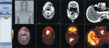
One condition that presents complications specific to each patient is obesity. According to the American Obesity Association, 20 percent of the U.S. population is obese, with six million of them morbidly obese. Consequently, these patients are hard pressed to get a CT scan due to the limited size of the CT’s bore. In response to this growing problem, GE Healthcare recently unveiled two new LightSpeed RT 16-slice CT models. The LightSpeed RT16 enables advanced imaging for radiation therapy, and the LightSpeed Xtra is a premium 16-slice CT scanner designed for interventional and bariatric procedures. The extra wide gantry opening and extended display field-of-view scans the entire anatomy of patients with this condition.
Both devices track tumor motion via integrated 4-D data support from new treatment planning platforms. “With 4-DCT we delivered a new standard of care in radiation oncology imaging of the chest, abdomen and pelvis; we have taken 4-D to the next level with the introduction of AdvantageSIM MD,” said Gene Saragnese, vice president and general manager of the company’s Global Molecular Imaging and CT business.
The Mind Behind the Machine
Behind every good modality is a solid software solution. “The precision of the software drives the hardware. You can upgrade hardware, but you have to have the proper software or you can’t be very successful,” explained Edward H. Gilbert, M.D., a radiation oncologist at the Richardson Regional Cancer Center in Richardson, TX. “It starts really with the software. Then if you take something like the Novalis – which really was invented for neurosurgery – the software and hardware design is so perfect that you can do very effective radiosurgery with the precision of 1 mm.”
To map out a treatment at submillimeter levels, treatment-planning software tools for IMRT, IGRT and SRS and SRT are critical to effectively radiating a moving target.
Optimizing the use of IMRT and IGRT in a software suite, both new LightSpeed scanners by GE are backed by Advantage 4D and Advantage SIM technology, fusing the capability of 4-D imaging and planning to deliver the highest levels of precision and speed for radiotherapy. Advantage 4D leverages the Varian Medical Systems RPM Respiratory Gating device, a system that allows the clinician to monitor and compensate for tumor movement throughout the process from simulation to treatment. “Advantage 4-D makes it possible to acquire CT scans that capture the motion of tumors and adjacent critical structures,” explained George T.Y. Chen, Ph.D., professor of Radiation Therapy at Massachusetts General Hospital.
In addition to the RPM Respiratory Gating solution, Varian also provides dynamic adaptive radiation therapy (DART) technology to support targeting in IMRT. The solution provides tools to manage inter- and intra-fraction motion and improve accuracy and performs both prospective and retrospective 4-D CT image acquisition.
The next step in Varian’s plan is to upgrade its current treatment planning system, Eclipse, to a 4-D system for gated respiratory motion throughout the entire treatment process. The system works with CT and PET/CT as well as pre-treatment fluoroscopy and DRR systems. “We are looking at the whole process from start to finish, adjusting the dose plan,” said Jefferson Z. Amaker, director, Treatment Planning Systems, Varian. “The concept of 4-D is for breathing and how the patient changes over the course of treatment.”
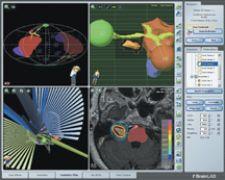
Streaming oncology into the future, BrainLab has introduced iPlan Flow, a new software for brain tumors that supports the planning of radiotherapy treatments in combination with local fluid delivery for chemotherapy, gene therapy, molecular-targeted therapy, immunotherapy and radioimmunotherapy. To complete the delivery of fluid infused into the interstitial space, a computer uses MR imaging to render an individual 3-D map of the patient’s brain. This map then calculates the distribution patterns of the medication, and with this information the physician can preoperatively identify the location to which the medication needs to be delivered.
“We can use this for all modalities, not just radiosurgery. Thus it provides an extension into our extracranial and IMRT treatments,” indicated Timothy Solberg, Ph.D., who is the professor and chief of Medical Physics at the University of Nebraska Medical Center, who has been using iPlan RT for several months. “We have also started to export the results of unique iPlan functions such as the atlas automatic organ segmentation into our existing RT planning system utilizing the DICOM RT standard.”
High-ART
Siemens recently outlined its blueprint for Adaptive Radiation Therapy (ART) solutions built on linac capabilities and brought it to market. MVision Megavoltage Cone Beam (MVCB) Imaging Package is a volumetric inline target imaging solution for IGRT. MVision employs the megavoltage (MV) source used for treatment to create a 3-D image of the patient.
Using a Siemens linear accelerator and standard flat-panel detectors, users can upgrade their existing technology with 3-D image-guided automated patient positioning software that is fully integrated in the workflow process. “ART is new to hospitals and users, and they are still learning about how to use it, which product to use and how often to use it on a patient,” noted Andreas Schlatter, director, Marketing Development, Siemens. “Working on protocols for ART as a community is very important.”
The backbone of the Elekta Synergy S, an image-guided robotic linear accelerator that combines high-conformance beam shaping for IGRT and stereotactic radiation treatments, is a 4-D adaptive IGRT technology for advanced stereotactic radiation treatments.
Integra Radionics recently upgraded its XKnife RT package for neurosurgeons, oncologists, physicists and dosimetrists to the 4.0 version. New features include sculpt mode contouring capability from inside or outside of the contour borders with a circular paint brush; bony segmentation to visualize bony anatomy without the need for contouring; split-slice contouring to visualize two correlated data sets simultaneously, while contouring on either data set; IGRT isocenter translations that automatically determine the isocenter translation needed to correct for patient changes when using CBCT or other daily imaging tools; and multiple isocenter translation to automatically restore the geometric relationship of each isocenter in the plan to the anatomy targeted during preplanning.
Drawing accurate conclusions is so critical to physicians, physicists and dosimetrists that Philips developed Pinnacle3 Model-Based Segmentation (MBS), a handy tool for contouring critical structure. The solution provides a library of predefined organ models. Users can drag and drop an organ model onto patient image data, and the software automatically adapts the model to fit the patient’s anatomy. The software also allows a facility to build its own library of organ models based on their own patients.
The art of better treatment planning involves better communication, and TomoTherapy attempts to bridge that gap with TomoGateway, a Virtual Private Network (VPN) connection that allows direct communication from each TomoTherapy Hi-Art System to the company’s contact center. The connection allows system data logs detailing the system’s operation and performance to be transferred to a customer support representative, eliminating the need for other reporting methods. It also allows technicians to perform online diagnostics and look at real-time data, while examining the system log files.
Six Degrees of Freedom
One of the technologies that has lifted stereotactic radiosurgery to new heights in adaptive radiotherapy is six degrees of freedom (6-D). 6-D is an image-guided robotic patient positioning system that corrects rotational errors in patient positioning as well as translational errors, enabling very high accuracy required for all radiation therapy, but in particular for stereotactic radiosurgery (SRS) and stereotactic radiation therapy (SRT).
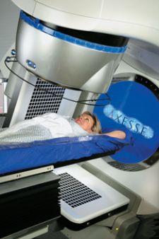
Elekta integrated 6-D into its Axesse system for radiosurgery of the spine as the free breathing delivery reduces treatment time and complexity, enables unique beam angle flexibility and highly conformal beam shaping.
The University of Pittsburgh Medical Center (UPMC), one of the leading radiosurgery centers in the U.S. and which has worked with Elekta to develop intracranial radiosurgery, recently implemented the Elekta Axesse system for extracranial radiosurgery. According to Anthony De Carolis, CEO of Elekta, Inc. and president of Elekta operations in North America, “Elekta Axesse is the most advanced system for image-guided stereotactic radiation therapy with the multifunction versatility required for extracranial applications.”
Earlier this year, specialists from MD Anderson put BrainLAB’s IGRT system – ExacTrac X-Ray 6D for lung and liver tumors – to the test. Through an integrated infrared tracking technology, the system acquires multiple stereoscopic X-ray images during the patient’s breathing cycle. By automatically calculating tumor movement in 3-D, the beams synchronize with the patient’s breathing frequency. If the tumor moves outside of the treatment area, radiation automatically freezes to protect surrounding healthy tissue.
“For the last 10 months we have used adaptive gating with ExacTrac X-Ray 6D to treat patients with lung tumors,” said Radiation Oncologist at MD Anderson Alan Forbes, M.D. “The adaptive gating technology reduces the area of the radiation treatment, which decreases the amount of lung tissue scarring and dose to critical structures. At the same time, the entire tumor target still receives the full dose of radiation. This allows me to implement more advanced treatment protocols, which will lead to better patient care at our center.”
Similarly, with robotic maneuverability and a full six degrees of freedom, CyberKnife claims that is has the ability to treat tumors from virtually any direction, both isocentrically and nonisocentrically.
In the quest for automated freedom, new robotic systems that use six degrees of freedom may one day enable physicians to move instruments for conformal radiation therapy exactly like their hand. Yet, while 6-D technology undergoes further clinical scrutiny, physicians will continue to rely on 4-D imaging to lead radiation therapy into the nearer future.