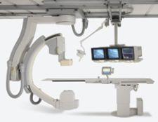
The Innova 4100 from GE Healthcare is the world's first large-format digital flat-panel X-ray system for angiographic imaging.
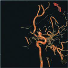
With an increased availability of effective imaging techniques, peripheral vascular disease diagnosis and treatment is continuously advancing. Procedures no longer rely solely on the seemingly archaic practice of using simple 2-D fluoroscopic images in the vascular lab to administer treatment. Interventional radiologists now have much more at their disposal. Some of the most notable areas of recent development have been in the interventional radiologists’ abilities to acquire 3-D at a digital flat-panel level of image quality in the vascular lab during the procedures. High-quality 3-D provides interventional radiologists with new tools that allow them to deliver safer, faster and more efficient therapy to the patient in real time.
Bringing 3-D Images into the Vascular Lab
Digital flat-panel 3-D imagery adds a new dimension of safety, planning and effectiveness to vascular procedures. It was a major breakthrough when an interventional radiologist was given the opportunity to acquire and visualize a 3-D magnetic resonance angiography (MRA) or computed tomography angiography (CTA) image to help guide and plan an intervention. But these images were acquired prior to the procedure, and if immediate 3-D images were needed, the patient had to be relocated from the vascular lab to the site of the CT or MRI systems.
This is all but a memory now. Today, interventional radiologists can acquire a 3-D image in the vascular lab, while the patient is on the table, in less than a minute in some cases. Currently, GE Healthcare, Philips Medical Systems, Siemens Medical Solutions and Toshiba America Medical Systems all offer more accessible 3-D capabilities.
“This is what we call rotational angiography, which is rotating the X-ray source and detector around the patient to collect what is essentially 3-D data sets,” said Brian Porras, director of product marketing, Interventional Radiology, Siemens Medical Solutions. “We can then take that data and not only select a 3-D data set, but take the next step and take that data set and reconstruct it into a cross-sectional image similar to what you would see on a CT scan.”
Quickly obtaining this CT-like 3-D image, while the patient is on the table in the lab, has several benefits. The interventional radiologist can see and measure the lesion from every direction without additional exposure or contrast media, which allows for better planning. If a patient is having a stent placed, for example, these images can be especially valuable.
“[Interventional radiologists] would perform measurements to determine the length and diameter of the lesion, and even potentially determine the type of stent that they were going to use,” said Karl Kellar, Americas vascular and interventional marketing manager, GE Healthcare. “Then, in real time, when they are inserting the device into the patient, they would have the [3-D] image, which can be used to guide the gantry to different viewing angles for fluoroscopy, and they could even perform another flat-panel CT image to make sure the deployment of that stent is correct after they put it in.”
Although 3-D capabilities are a tremendous help to the interventional radiologist for treatment planning, the real-time 2-D fluoroscopic image is still used in the vascular lab to guide the radiologist in real time while performing an internal procedure on the patient.
On the Verge of Real-Time 3-D Images
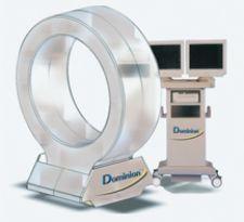
There is however, a blossoming technology that does make it possible to view 3-D images in real time. Imaging3 Inc., a company based in Burbank, CA, has received the patent for a proprietary medical technology called Dominion Technology, which will produce 3-D medical diagnostic images in real time. This means that physicians can instantly view high-resolution 3-D images of any desired area of the body.
“The Dominion will take a really big step up in terms of the accuracy for surgical navigation and so on,” said Mike Nessen, vice president of Business Development, Imaging3 Inc. “What the Dominion does in its first revolution, which takes eight-tenths of a second, is orient the body and find out where it is in relationship to the gantry. Each time it images it is reorienting itself to the body, which can get down to micron-level accuracy. There is definitely an accuracy advantage.”
While other systems are capturing 3-D images in around a minute or longer, the Dominion’s ability to allow the radiologist to view a 3-D image in two-tenths of a second could revolutionize peripheral vascular interventional procedures. Moreover, the system is mobile, which will allow its multimodality CT, fluoroscopy and real-time 3-D capabilities to be used in multiple capacities including the ER and OR, in addition to vascular labs. Imaging3 will demonstrate this technology for the world to see at the Radiological Society of North America’s 92nd Scientific Assembly and Meeting in Chicago Nov. 26 - Dec. 1, 2006.
Possibilities of 2-D/3-D Integration
At this juncture, the interventional radiologist can acquire the 3-D image of the area of interest and import it into the workstation while the patient is on the table to use as a guide. Right next to that monitor is the real-time 2-D fluoroscopic image for therapeutic purposes. Now, what if it were possible to superimpose that 3-D image over the 2-D fluoroscopic image for the “ultimate” therapeutic tool? This 2-D/3-D integration is a technique that may not be too far-fetched.
This technology is currently being developed and validated, and may be something that becomes commonly available over the next couple of years.
“[2-D/3-D integration] would allow for the interventionalist to have a better feel of where they are in [the patient] anatomically, and it would overlay the anatomical information right over the live fluoro as an additional image-guidance capability to help guide the therapy,” stated Porras.
Kellar noted that MR and conventional CT images do a better job at imaging soft tissue at the present time. Therefore, if they could be superimposed with the high-detail X-ray rotational 3-D images, which show more detail in the high-contrast vessels, the interventional radiologist would be able to see where the fine detail vascular circulation is compared to the surrounding tissue of the 3-D image from other modalities.
Another area that could see advantages of the superimposition is catheter placement. A 3-D component to the live 2-D fluoroscopic image would help the interventional radiologist more accurately steer the catheter in the exact direction intended.
“As one could imagine, there is a lot of real-time calculation inherently involved in this, and there is some good research which has proven we can do the math, and we can accurately do the superimposition,” added Kellar. “So over the next few years, this is going to become a useful navigation aide for people that are actually steering the catheter itself.”
Easy-Access IVUS
Just as 3-D imaging is a growing technology in the world of peripheral vascular imaging, so is intravascular ultrasound (IVUS), which can be particularly useful for viewing and analyzing plaque. IVUS has been around for a number of years, but only now is it becoming convenient enough for routine use. It used to be IVUS systems were bulky machines that were brought into the vascular lab as needed and set up for use. This took significant time and required the room to be reconfigured to accommodate the IVUS system. GE Healthcare integrates the Volcano IVUS system into the tableside controls of their Innova digital flat-panel X-ray systems for instant access to IVUS in the vascular lab without bringing in additional hardware.
“We have integrated the IVUS system into our tableside controls so that literally, all the doctor has to say is ‘I would like to get an IVUS on this patient’, select the appropriate IVUS catheter, plug it in, and get the information in a matter of moments and be done,” said Kellar.
The addition of these exciting new technologies to the treatment of peripheral vascular disease has the potential to increase accuracy of real-time decision- making, speed up procedures and reduce the amount X-ray dose and contrast materiel used. These benefits are good for both the patient and physician alike and represent a real advance in image-guided intervention.