June 14, 2023 — A University of Waterloo engineer’s MRI invention reveals better than many existing imaging technologies how COVID-19 can change the human brain.
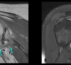
The new imaging technique known as correlated diffusion imaging(CDI) was developed by systems design engineering professor Alexander Wong and recently used in a groundbreaking study by scientists at Baycrest’s Rotman Research Institute and Sunnybrook Hospital in Toronto.
“Some may think COVID-19 affects just the lungs,” Dr. Wong said. “What was found is that this new MRI technique that we created is very good at identifying changes to the brain due to COVID-19. COVID-19 changes the white matter in the brain.”
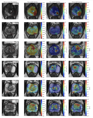
Wong, a Canada Research Chair in Artificial Intelligence and Medical Imaging, had previously developed CDI in a successful search for a better imaging measure for detecting cancer. CDI is a new form of MRI that can better highlight the differences in the way water molecules move in tissue by capturing and mixing MRI signals at different gradient pulse strengths and timings.
Researchers at Rotman, a world-renowned center for the study of brain function, saw Wong’s imaging discovery and thought it could likely also be used to identify changes to the brain due to COVID-19. Subsequent tests proved that theory right. The CDI imaging of frontal-lobe white matter revealed a less restricted diffusion of water molecules in COVID-19 patients. At the same time, it showed a more restricted diffusion of water molecules in the cerebellum of patients with COVID-19.
Wong highlights that the two regions of the brain react differently to COVID-19 and points to two key findings from the research. First, the human cerebellum might be more vulnerable to COVID-19 infections. Second, the study reinforces the idea that COVID-19 infections can lead to changes in the brain.
Not only is the Rotman study one of the few to have shown COVID-19’s effects on the brain, but it is the first to report diffusion abnormalities in the white matter of the cerebellum. Although the study was designed to show changes, rather than specific damage, to the brain from COVID-19, its final report does discuss potential sources of such changes and many link to disease and damage.
In response, Wong suggests future tests could focus on whether COVID-19 actually damages brain tissue. Additional studies could also determine if COVID-19 can change the brain’s grey matter.
“Hopefully, this research can lead to better diagnoses and treatments for COVID-19 patients,” Wong said. “And that could just be the beginning for CDI as it might be used to understand degenerative processes in other diseases such as Alzheimer’s or to detect breast or prostate cancers.”
The study, Feasibility of diffusion-tensor and correlated diffusion imaging for studying white-matter microstructural abnormalities: Application in COVID-19, which involves Wong and his student Hayden Gunraj as co-authors, is published in the journal Human Brain Mapping.
For more information: https://uwaterloo.ca/
Related content:
MRI Reveals Significant Brain Abnormalities Post-COVID
MRI Innovation Makes Cancerous Tissue Light Up and Easier to See
Long COVID Syndrome in Children and Teens
Long COVID Implications: Increased Health Care Use After Infection With SARS-Cov-2
Lasting Lung Damage Seen in Children and Teens after COVID
PHOTO GALLERY: How COVID-19 Appears on Medical Imaging
COVID-19 Fallout May Lead to More Cancer Deaths
Kawasaki-like Inflammatory Disease Affects Children With COVID-19
FDA Adds Myocarditis Warning to COVID mRNA Vaccine Clinician Fact Sheets
CMS Now Requires COVID-19 Vaccinations for Healthcare Workers by January 4
Cardiac MRI of Myocarditis After COVID-19 Vaccination in Adolescents
Small Number of Patients Have Myocarditis-like Illness After COVID-19 Vaccination
Overview of Myocarditis Cases Caused by the COVID-19 Vaccine
Case Study Describes One of the First U.S. Cases of MIS-C
NIH-funded Project Wants to Identify Children at Risk for MIS-C From COVID-19
 May 08, 2024