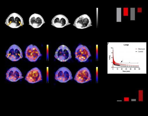
A) Axial CT images through the mouse lungs at 7 and 14 days after intratracheal administration of bleomycin or saline (as a control), demonstrating increased lung fibrosis in the bleomycin group (white arrows). (B) CT attenuation histograms in Hounsfield units (HU) after lung segmentation demonstrate increased attenuation in the lungs in the bleomycin group than the control group (p <0.05), consistent with increasing fibrosis (n=3). (C) Representative axial PET/CT fusion images at 20 and 60 min demonstrating increased FAPI uptake in the lungs of the bleomycin group (white arrows) with no significant uptake in the control group (yellow arrows). (D) Time-activity curve of lung uptake ROI analysis demonstrating higher FAPI uptake in the lungs of the bleomycin group than the control (p < 0.05), 14 days after bleomycin (n=3). (E) Ex vivo biodistribution data of lung tissue demonstrating higher radiotracer uptake in the lungs of the bleomycin group than the control (n=3). *p<0.05, **p<0.01. Image created by CA Ferreira et al., University of Wisconsin-Madison, Madison, WI.
June 14, 2021 — Positron emission tomography (PET) using a 68Ga-labeled fibroblast activation protein inhibitor (FAPI) can noninvasively identify and monitor pulmonary fibrosis, according to research presented at the Society of Nuclear Medicine and Molecular Imaging 2021 Annual Meeting. By binding to activated fibroblasts present in affected lungs, FAPI-PET allows for direct imaging of the disease process.
Idiopathic pulmonary fibrosis (IPF) causes substantial scarring to the lungs, making it difficult for those impacted to breathe. It is a significant cause of morbidity and mortality in the United States, with more than 40,000 deaths annually. A major challenge in diagnosis and treatment of IPF is the lack of a specific diagnostic tool that can noninvasively diagnose and assess disease activity, which is crucial for the management of pulmonary fibrosis patients.
"CT scans can provide physicians with information on anatomic features and other effects of IPF but not its current state of activity. We sought to identify and image a direct noninvasive biomarker for early detection, disease monitoring and accurate assessment of treatment response," said Carolina de Aguiar Ferreira, PhD, a research associate at the University of Wisconsin-Madison in Madison, Wisc.
In the study, researchers targeted the fibroblast activation protein (FAP) that is overexpressed in IPF as a potential biomarker. Two groups of mice--one group with induced pulmonary fibrosis and one control group--were scanned with the FAPI-based PET/CT radiotracer 68Ga-FAPI-46 at multiple time points. Compared to the control group, the mice with induced pulmonary fibrosis had a much higher uptake of the radiotracer, allowing researchers to successfully identify and evaluate areas of IPF.
"Further validation of 68Ga-FAPI-46 for the detection and monitoring of pulmonary fibrosis would make this molecular imaging tool the first technique for early, direct, and noninvasive detection of disease. It would also provide an opportunity for molecular imaging to reduce the frequency of lung biopsies, which carry their own inherent risks," noted Ferreira. "This development will demonstrate that functional imaging can play an invaluable role in evaluation of the disease process."
Abstract 10. "Targeting Activated Fibroblasts for Non-invasive Detection of Lung Fibrosis," Carolina Ferreira, Zachary Rosenkrans, Ksenija Bernau, Jeanine Batterton, Christopher Massey, Alan McMillan, Nathan Sandbo, Ali Pirasteh and Reinier Hernandez, University of Wisconsin - Madison, Madison, Wisconsin; and Melissa Moore, Frank Valla and Christopher Drake, Sofie Biosciences, Dulles, Virginia.
For more information: www.snmmi.org

 April 29, 2024