Feature | December 13, 2013

System can identify chemical composition of materials in the body

spectral CT, IQon

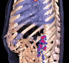
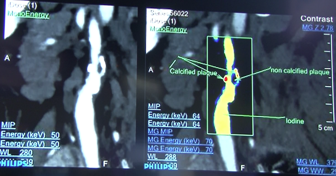
Philip's spectral CT showing how the system can identify the atomic number of iodine and calcium from photons on the scan and overlay a color map showing the chemical composition of materials in the body. This image shows a traditional grayscale Hounsfield image of a vessel and the right image showing a spectral version showing color-coded iodine and calcium.


December 13, 2013 — Philips Healthcare introduced the IQon Spectral computed tomography (CT) system at RSNA 2013. It is the world’s first spectral-detector CT system built from the ground up for spectral imaging. It uses color to identify the composition of an image without involving time-consuming protocols. 
 
In the same way that white light is made up of a spectrum of colors, the X-ray beam photons used in CT scanners also consist of a spectrum of X-ray energies. With the development of a fundamentally new spectral detector that can discriminate between X-ray photons of multiple high and low energies simultaneously, Philips’ IQon Spectral CT adds a new dimension to CT imaging, delivering not only anatomical information but also the ability to characterize structures based on their material makeup within a single scan. 
 
Using a type of spectral analysis, the system can separate out materials made up of specific atomic numbers off the periodic chart of elements. The system so far has been tested for iodine and calcium. This can be used to help differentiate between areas of high blood contrast uptake and calcified areas, which can be useful in diagnosing kidney stones and better delineating various types of atherosclerotic plaque in arteries. Elements can be assigned specific color codes to make them standout on scans, even if the surrounding tissue has similar Hounsfield unit numbers. 
 
After a spectral CT examination, clinicians can interpret the conventional grey-scale anatomical images, and if necessary, access the spectral information that was acquired during the same scan. The IQon Spectral CT system’s retrospective on-demand data analysis is made possible via Philips’ iPatient platform, allowing clinicians to easily experience the benefits of spectral CT routinely within traditional radiology workflows. 
 
Due to its accessibility, speed and accuracy, CT imaging is widely used in the diagnosis of many different diseases and injuries, totaling approximately 450 million imaging procedures globally per year. Philips has consistently driven innovation in CT, most recently with the introduction of its proprietary Iterative Model Reconstruction (IMR) technology to simultaneously reduce CT radiation dose4 and enhance image quality for a broad range of applications. 
 
The IQon Spectral CT is pending 510(k) and is not available for sale in the United States. A system with the NanoPanel Prism detector is available for sale in the United States. for conventional scanning.
 
For more information: www.philips.com/newscenter


Related Content

Technology

Swissray’s ddRCruze provides increased flexibility for digital radiography (DR) systems at modern healthcare facilities. The vendor asked technologists to help design the mobile X-ray system.

Time October 17, 2014
arrow
Technology

Invivo has expanded its breast and prostate imaging solutions portfolio with the addition of the Sentinelle product line of advanced MRI coils.

Time October 13, 2014
arrow
News

GE Healthcare, a unit of General Electric Company, developed features on the Logiq S8 general imaging ultrasound system. The innovations allow for enhanced image quality and streamlined workflow for healthcare providers, and help shorten exam times for patients. GE presented the advancements at the Radiological Society of North America (RSNA) 2013 conference.

Time February 03, 2014
arrow
Feature | Raissa Rocha

The practice of breast imaging and screening in women at risk for breast cancer is evolving as new imaging modalities are incorporated and researchers continue to study the implications of dense breast tissue in patients. At the 2013 annual meeting of the Radiological Society of North America (RSNA) in Chicago, there were several sessions highlighting trials and studies that brought breast density and cancer detection to the forefront.

Time February 03, 2014
arrow
Feature | Williette Nyanue

Several industries have used cloud solutions for many years, but cloud computing only recently started to be used in healthcare. According to the National Institute of Standards and Technology (NIST), cloud computing is defined as “a model for enabling convenient, on-demand network access to a shared pool of configurable computing resources (e.g., networks, servers, storage, applications and services) that can be rapidly provisioned and released with minimal management effort or service provider interaction.”1 As more and more healthcare organizations (HCOs) adopt electronic medical records (EMRs), the cloud database has offered an efficient solution for image sharing, particularly in radiology where it is bridging the gap between referring physicians and radiologists.

Time February 03, 2014
arrow
Feature | Greg Freiherr

What was once diagnostic imaging is on track to becoming much more. Radiology is on the verge of a new era in which its focus goes beyond the traditional gatekeeper role, directing the earliest steps in patient management to one that adjusts the direction of care throughout the management of a patient.

Time February 03, 2014
arrow
Feature | Williette Nyanue

Physicians have used radiation in medicine for more than a century. The use of radiation in diagnostic imaging, including computed tomography (CT), fluoroscopy, angiography, mammography, computed radiography (CR) and digital radiography (DR), as well as in nuclear medicine, has aided greatly in the diagnosis and treatment of cancer and other diseases.

Time February 03, 2014
arrow
News

RamSoft’s PowerServer 6.0 features updated aesthetics to aid usability.

Time January 20, 2014
arrow
Technology

Sony Electronics’ Medical Systems Division unveiled at the 2013 Radiological Society of North America (RSNA) meeting its next generation of medical printers, including three digital thermal models with features such as built-in image capture and hybrid signal inputs.

Time January 20, 2014
arrow
News

Sony Electronics’ Medical Systems Division demonstrated its latest developments in 4K and 3D imaging technologies designed to re-shape visualization, training and education for a range of medical applications.

Time January 20, 2014
arrow
Subscribe Now