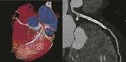
Coronary CTA on the Somatom Force produces high-resolution stent imaging with a 0.18 second scan at 70 kV exposing the patient to just 0.43 mSv. The patient's heart rate varied between 58 to 70 bmp during the exam. Color image shows two long stents. The curved MPR image in grayscale shows the details in the LAD stent. Image provided by Siemens Healthcare, copyright Institut fur Klinische Radiologie und Nuklearmedicin, Universitatsmedizin Mannheim Medizinische Fakultat Mannheim der Univesitat Heidelberg
What was once diagnostic imaging is on track to becoming much more. Radiology is on the verge of a new era in which its focus goes beyond the traditional gatekeeper role, directing the earliest steps in patient management to one that adjusts the direction of care throughout the management of a patient.
The cornerstone of this new era — patient-centric imaging — was the spark behind last year’s “Patients First” campaign by the Radiological Society of North America (RSNA), which, although out of sync with the reality of radiological practice, was correct at its core in that radiology must find a way to increase its relevance to the patient. It came into tune this year, when the best interests of patients aligned with those of radiologists, not on placards dotting the hallways of the annual meeting, but in new unveilings.
Examples of transformative technologies were seen on the exhibit floor of RSNA 2013. Siemens brandished a new magnetic resonance (MR) technology, called FREEZEit, that promises to vanquish motion artifact from liver images. Physicians seeking insights into these very sick patients must now rely on contrast-enhanced computed tomography (CT) images. FREEZEit would provide a gentler alternative.
A new generation of CT scanners is being readied to produce angiograms, captured at high speeds and paradoxically low radiation dose, that might allow the more accurate planning and effective implantation of stents, as well as repair heart valves; for example, transcatheter aortic valve implantations. Such exams are possible now only with Toshiba’s pathfinding Aquilion One, but soon competitors from Siemens and GE, shown as works in progress at this year’s RSNA meeting, will enter the market.
The GE Revolution CT can capture a human heart in a single beat free from motion artifact. At the show, the company framed the 510(k)-pending system as an “all-in-one” scanner, offering coverage, spatial and temporal resolution that can handle a comprehensive range of radiological applications, while serving as a platform for new applications.
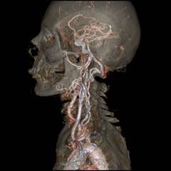
Siemens billed its Somatom Force as the next generation in dual-source CT. Successor to the Somatom Definition Flash, the company’s previous top-tier CT, Force features two generators that — like the generators on its predecessor — fires X-rays independently at variable energies. Force is so fast, however, that it can image the adult chest in a second, eliminating the need to breathhold, and freeze a human heart pumping at 90 beats per minute by merging the two imaging chains to create a 50 cm (nearly 19.7-inch) field of view. Alternatively the two imaging chains, like those on the Flash, can be tuned to separate energies to differentiate among tissues or materials, as in visualizing iodinated contrast as it flows through the coronaries or accumulates in varying amounts in benign versus malignant masses, such as those in the kidney.
Newly unveiled at RSNA 2013 and 510(k)-pending, the IQon Spectral CT from Philips attempts such differentiation using a single imaging chain to capture X-rays at different wavelengths. Colors identify the composition of structures. Cancer appears bright red due to a concentration of iodinated contrast medium in the vascular bed feeding a tumor. A pulmonary embolism appears in blue due to a lack of vasculature. Conventional gray scale images show the underlying anatomy.
Returning to RSNA after an extended hiatus, SonoSite reappeared this year as a component of Fujifilm. The pioneer of hand-carried ultrasound did so with what appeared as a cart-based system, the X-Porte. Company reps demurred, referring to X-Porte as a “kiosk,” a deserved distinction given the product’s novel touchscreen interface, featuring four large, rectangular, multi-colored soft buttons — green, blue, purple and red — whose functions can be customized to operator preferences. As with its hand-carried brethren, the new system is designed for point-of-care, allowing easy transport within a hospital or clinic, say advocates. But X-Porte goes beyond them with a new imaging architecture, called Extreme Definition Imaging, and a beamforming algorithm that improves image quality. Just as hand-carried systems can be overnighted to SonoSite for immediate replacement, the engine that drives X-Porte — about the size of an Xbox — pops out for quick mailing and return, in the event of failure.
Supersonic Imagine made the jump to mainstream releasing at the show the Expert edition of its Aiexplorer ultrasound platform, a cart-based scanner newly plied with an OB/GYN application that filled the previous gap in radiological capabilities. No longer constrained by a lack of applications, the Expert Edition Aixplorer had made the leap from a specialty unit for elastography to a premium radiology platform, featuring superfast color Doppler and elastography for breast, liver and thyroid — elastography that,
with the acceptance of the U.S. Food and Drug Administration (FDA), rendered quantitative numbers.
Quantitation — A Key Enabler
In radiology’s transformation to purveyor of total patient management, quantitation will be a key enabler. Quantitation turns subjective observations into objective measures upon which radiologists and their referring physicians can base patient management decisions.
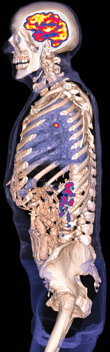
Siemens shone a spotlight on its Biograph mCT Flow PET/CT scanner for improvements in image quality and standard uptake values (SUV) thanks to a continuously moving patient table that ushers body areas steadily through the detector, producing a more reproducible and robust stream of data. The company’s Symbia Intevo single-photon emission computed tomography (SPECT)/CT leveraged the CT data set to create more accurate SPECT images, as well as combining the two data sets to allow, for the first time, accurate quantitation on a SPECT system.
What mCT Flow could do at the premium end of molecular imaging, Symbia Intevo might do for the masses performing bread-and-butter nuclear scans. Early adopters will see the benefit in bone scans, as Siemens has honed the technology first for this application. Other applications are expected to follow.
Philips further extended the power of positron imaging with a solid-state replacement for the photomultiplier tubes that have registered photons in PET scanners since their development in the 1970s. Philips positioned the new detector as the core of the industry’s first truly digital PET/CT, the company’s new Vereos. The new system, already cleared by the FDA, is truly remarkable as a pathfinder technology and a watershed development for Philips, which was late to market with a PET/CT but now may have an edge, as the PET installed base prepares to replace aging systems. The company claims its detector produces images with two times better image resolution than PMT-based scanners and SUV measurements that are twice as accurate.
Also standing to reap the benefits of quantitation is women’s health. More than a dozen states have enacted legislation requiring physicians to inform patients with dense breasts that mammography may not effectively pick up the earliest signs of cancer. Here quantitative measures of breast density may determine whether tests other than mammography should be employed as a screening tool.
In this Philips has the ideal detector, a photon-counting technology that underlies its digital MicroDose mammography system. Complementing its quantitative abilities is the decidedly low-dose character of Philips breast system, which the company purchased two years ago from Sectra.
Safety and Comfort
Visitors to the RSNA exhibit floor heard a familiar mantra — low dose imaging — mouthed by the vendors of all things ionizing: CT, radiography, fluoroscopy/angio, mammography and molecular imaging. A fluoro feature for Toshiba’s Infinix-i cardiovascular systems tantalized for its simple, yet novel approach to minimizing dose. By their nature, interventional procedures demand real-time imaging. Toshiba’s Spotfluoro minimizes radiation exposure by surrounding a rectangular region of interest (ROI) with a “last-image hold,” an image captured some seconds earlier. Updating the image in the ROI exposes the patient to less dose, just as it keeps the interventionalist current on what’s happening.
The three premium CT scanners unveiled by GE, Siemens and Philips at RSNA each addressed low-dose scanning. GE’s Revolution CT features ASiR-V, a hybrid of the company’s iterative reconstruction software, called ASiR, and its model base, Veo. Philips’ proprietary iterative modeling technology, IMR, built for the company’s CT line, was featured on its newly unveiled IQon Spectral CT.
The two X-ray generators on Siemens’ Somatom Force can be pumped at low voltage (as low as 70 kVp) to deliver low-dose scans. Dose can be reduced further using the company’s SAFIRE software. This combination allows Somatom Force to scan the lungs at half the dose of comparable systems, according to Siemens, making this 510(k)-pending technology ideal for lung cancer screening, which could soon enter mainstream medical practice in the United States.
Over the years, Philips, GE and to some degree Toshiba have dabbled in ways to make the patient environment less plain and institutional and, thereby, more comforting. They have proposed — and sold to some sites — patient suites comprised of calming colors, scents and pastoral viewscapes. Such non-opiate relaxants were the key to GE’s Sensory Suite, shown through virtual goggles and in Jetson-like chairs in the GE booth.
Noise pollution, a problem in the MR suite since the very beginning of this modality, has been addressed mostly through the use of squishable ear plugs, handed out by techs. Wedge those chunks the wrong way and the bizarre noises that characterize MRI are a patient’s constant — and very loud — companions. Last year GE unveiled Silent Scan, a partial solution to the problem for one MRI sequence, a solution the company expanded this year to four more sequences. While Silent Scan is anything but silent, it helps. So much, in fact, that Siemens followed suit with its QuietSuite, which — like SilentScan — reduces the noise made by the body coil that surrounds the patient.
Increased Relevance, Enhanced Value
By becoming deeply embedded in patient management and making their reports and consultation of greater value to referring physicians, the relevance of radiologists will naturally increase. As the radiology community begins this transformation, they have begun to redefine productivity from more cases to cases reported more quickly.
Seeking efficiencies and lower costs, Viztek moved its picture archiving and communication system (PACS) platform to the cloud with Web-based technologies that support PACS functions and display images on both fixed and mobile devices. This new PACS works on Windows, MAC and Android devices, allowing flexible and secure reads.
Carestream Health came to RSNA 2013 with a work in progress Vue for teleradiology PACS module designed to support off-site reading and reporting by radiology group practices with multiple clients. The vendor neutral technology leverages the company’s Vue Connect platform to access off-site worklists, automatically delivering reports and immediately alerting referring docs to their availability. Built into those reports, using Carestream’s Vue Reporting, are embedded hot links that, when clicked, snap the reader to key images embedded in the report and arrows pointing out pathologies.
Softek featured the latest version of Illuminate’s PatientView, which pulls clinical notes from the EMR, displaying them along with prior radiology, pathology and lab reports. Information appears in a window that automatically opens when the radiologist pulls up a study.
Efficiency demands automation, and radiologists have been the beneficiaries of such automation for
the better part of two decades. Algorithms increasingly have done away with the knob-twisting in modalities as diverse as ultrasound and MRI, producing reproducibly high-quality images with ever less dependence on operator skill. In the future, efficiency will be characterized by automation that works in novel ways.
Samsung’s work in progress UGEO WS80A does that in five dimensions. The 5-D moniker is more marketing than real, although “5-D imaging” rightly indicates a new approach. In this, volumetric data acquired by the tech are plumbed for the relevant views. In the case of a fetal femur, for example, this bone is measured as well as displayed immediately after the operator requests results for that specific OB/GYN exam.
Such novel ways of boosting efficiency are all the more important now that radiologists are angling to contribute to the whole of patient management. Value is the common denominator of the new state of radiological relevance — and the means to relevance is making the most of radiologists’ special skills, enhancing those skills with technology that reveals better quality information and distributing radiologists’ interpretations quickly and effectively.
The seeds of this revolution were sown in the form of systems debuting at RSNA 2013. Some are months away from being commercially available. It may take even longer for them to be widely embraced. But the wheels are in motion, as radiology appears headed for an interesting and new phase.

 April 17, 2024


